
Gallbladder Abscess
Copyright 2008
Alok Anand Ashley Davidoff MD
Definition
Gallbladder abscess is an advanced form or complication of acute cholecystitis caused by a progressive inflammatory/infectious process that spreads to the gallbladder fossa and sometimes to the free wall of the gallbladder. The wall breaks down either due to ischemic necrosis or due to destruction by the infectious process, and fluid, necrotic tissue, viable and non viable white cells, viable and non viable bacteria, and tissue debris accumulate and are surounded by a wall of inflammed tissue.
Structurally the matrix of the abscess is of a complex fluid nature, is of variable size, is round and under pressure, and is confined by a wall of inflammed surrounding tissue.
Functionally in and of iteself it does not cause significant functional impairment, but the systemic effects of severe inflammation result in fever, malaise, anaorexia, and tiredness which are reflected in the clinical examination.
The diagnosis is confirmed by imaging techniques, usually ultrasound ,which is the procedure of choice in patients with right upper quadrant ultrasound.
CTscan and MRI are usually helpful in providing a global view of the extent of the disease.
Treatment requires both decompression of the obstructed gallbladder and the abscess and is best performed by percutaneous image guided techniques. This is usually followed by elective cholecystectomy when the patient is in better condition.
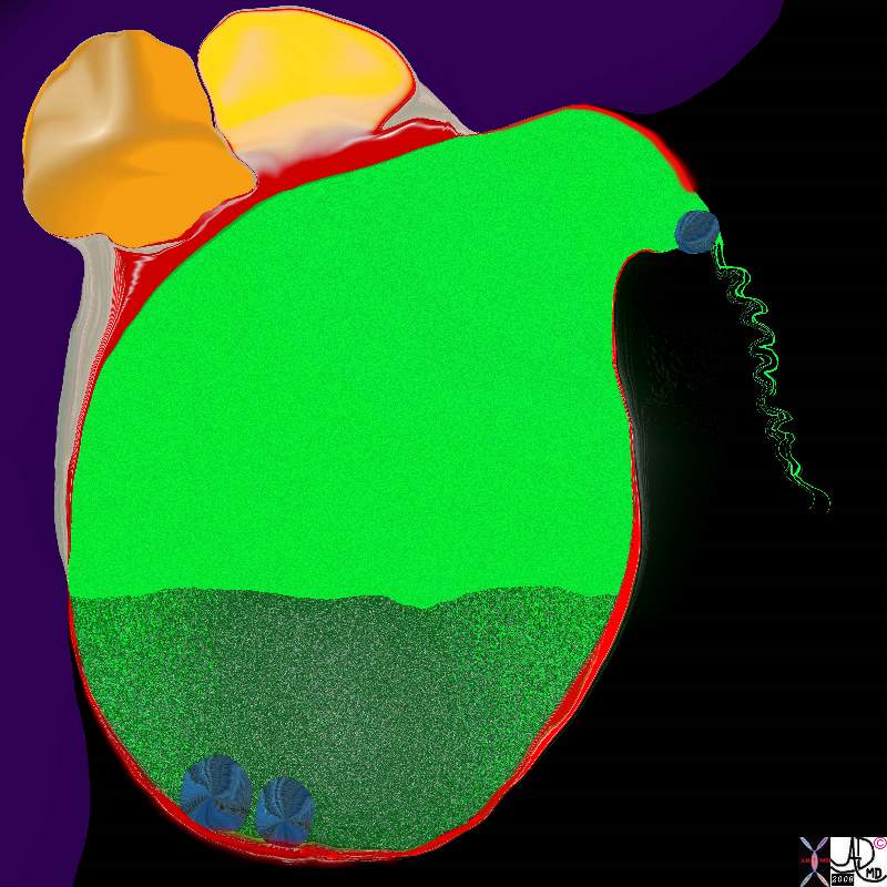
Cholecystitis Complicated by Abscess Formation in the Gallbladder Fossa |
|
The diagram illustrates the findings of acute cholecystitis caused by a stone impacted in the neck of the gallbladder, with secondary distension of the gallbladder and inflammation of the wall. (red). The thickened sediment represents gallbladder sludge and is caused by ongoing cholestasis. The infected bile and inflammatory process has advanced through a necrotic wall and into the gallbladder fossa where a a biloculate pus filled abscess cavity has formed. 11921.8b05b037.8s gallbladder cystic duct gallstones cholelithiasis stone impacted in the cystic duct distended enlarged hyperemic wall complex fluid in the gallbladder fossa tumefactive bile cholestasis sludge acute cholecytitis abscess formation Davidoff Art copyright 2008 |
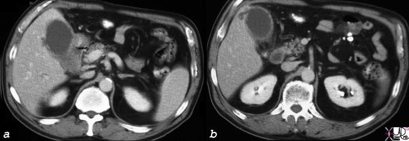
Early Abscess Formation |
|
The CTscan is from a patient who has a fever and right upper quadrant pain and who has acute cholecystitis. A small fluid collection is seen in the gallbladder fossain both images a and b that reflects early abscess formation. 16188c.8s gallbladder hyperemic thickened wall early abscess formation fluid collections in the gallbladder fossa acute cholecystitis CTscan Courtesy Ashley Davidoff MD copyright 2008 |
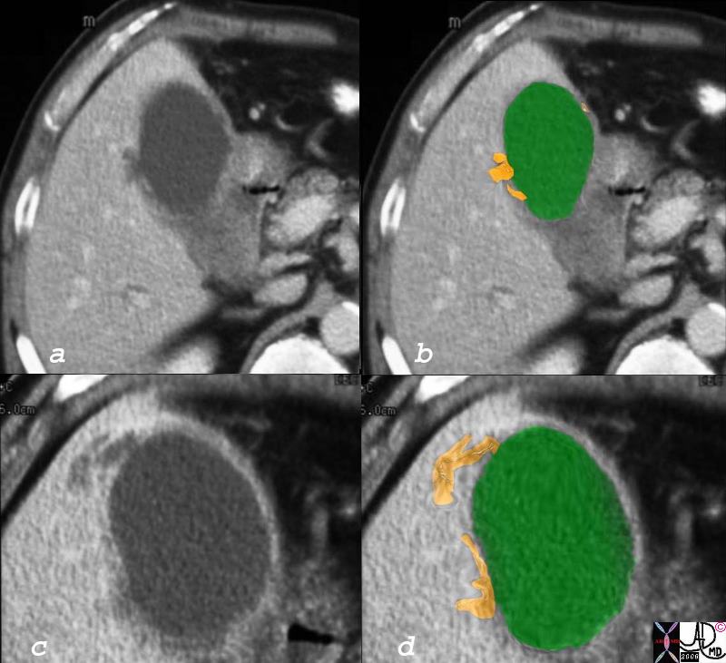
Early Abscess Formation |
|
The CT scan through the gallbladder shows a second case of early spread of the infection into the gallbladder fossa. The yellow more linear collections represent the early purulence which at this stage are too small for drainage and a trial of antibiotics would be the treatment of choice. 16188c05.8 gallbladder hyperemic thickened wall early abscess formation fluid collections in the gallbladder fossa acute cholecystitis CTscan Courtesy Ashley Davidoff MD copyright 2008 |
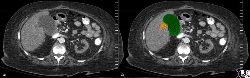
Abscess in the Gallbladder Fossa |
|
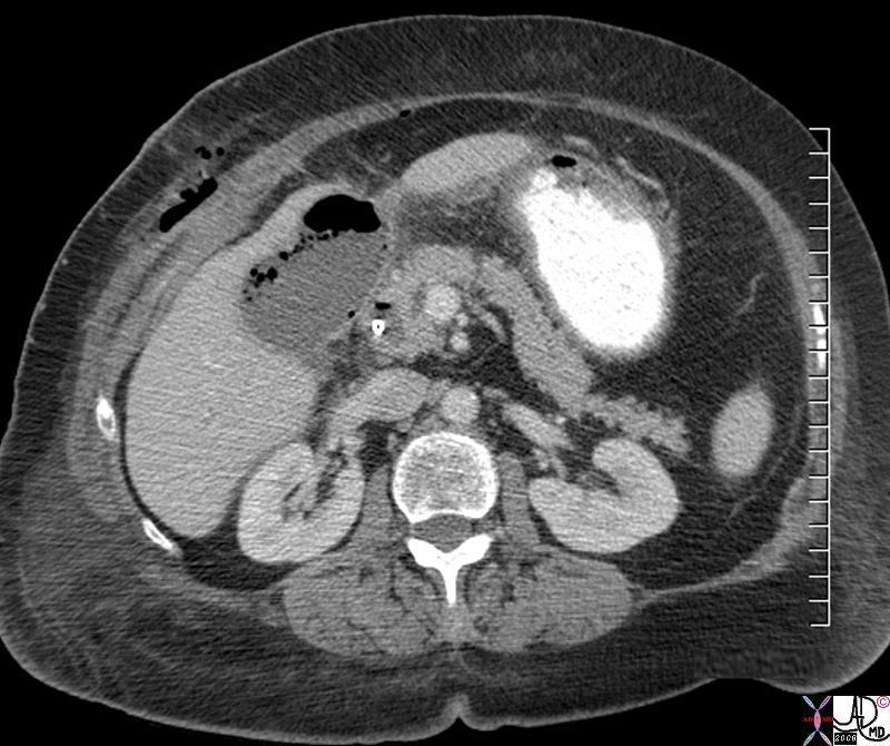
In this CTscan through the gallbladder fossa the abscess is becoming more confluent, and rounded but is still relatively small. One could consider a combination of aspiration, saline flush and antibiotics as the treatment in this case. The collection is too small for catheter drainage, since the pigtail would not be able to form in the collection and drainage would be suboptimal. 77673c02.8s elderly female with RUQ pain and fever gallbladder distended complex fluid collection with hyperemic rim in gallbladder fossa dx abscess in gbf secondary to acute cholecystitis Courtesy Ashley DAvidoff MD copyright 2008 |
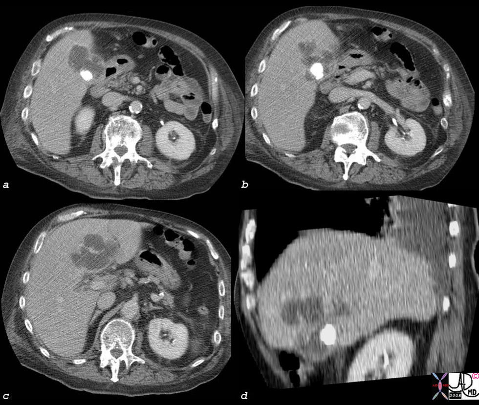
Abscess in the Gallbladder Fossa |
| The abscess in the gallbladder fossa in this patient is large and irregular and is associated with a large calcified stone in the gallbladder. Images a,b,c, are in the transverse plain, while image d is in the coronal plane. It is in the latter situation, where the stone can be seen in the relatively small gallbladder and the abscess in the gallbladder fossa and within the liver is optimally seen. percutaneous drainage and antibiotics is indictated. In this instance gallbladder carcinoima should alsd be considered in the appropriate clinical setting.
44364c02s elderly patient with fever right upper quadrant pain, RUQ pain, elevated white cell count gallbladder cholelithiasis acute cholecystitis fluid in the gallbladder fossa gallbladder abscess liver abscess hepatic abscess CTscan ‘Courtesy Ashley Davidoff MD copyright 2008 |
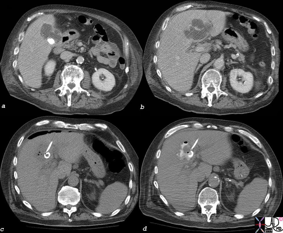
Abscess in the Gallbladder Fossa Treated with Catheter Drainage |
|
The patient above was treated with percutaneous drainage, (c) was decompressed overnight and the abscessogram performed the following day shows contrast in a decompressed abscess. Drainage should continue till the cavity is less than 5-100ccs in volume and the drainage is less than 5-10ccs over 24 hours. 44364c03s elderly patient with fever right upper quadrant pain, RUQ pain, elevated white cell count gallbladder cholelithiasis acute cholecystitis fluid in the gallbladder fossa gallbladder abscess liver abscess hepatic abscess percutaneous drainage catheter drainage MIT minimally invasive therapy CTscan ‘Courtesy Ashley Davidoff MD copyright 2008 |
Abscess in the Gallbladder Fossa s/p cholecystectomy |
|
The CTscan represents an abscess in the post operative bed of a patient who had undergone cholecystectomy. The post op fossa and abscess has the same pyriform shape as the gallbladder and contains a fluid collection with bubbles of air characteristic of an infection with anerobic organisms. the bubbles although floating to the top are not quite free to form an air fluid layer indicating complex fluid probably with thick pus and fibrinous strands. Percutaneous drainage is indicated. of a different type 77668.8s gallbladder gallbladder fossa s/p cholecystectomy skin bile duct stent air fluid air fluid level s/p cholecystectomy abscess in post operative bed CTscan Courtesy Ashley DAvidoff MD copyright 2008 |