
The Common vein Copyright 2008
Definition
Adenomyomatosis is a hyperplastic disorder of the mucosa, The cause is uncertain, but it has been linked with increased intraluminal pressure in the gallbladder, and to hyperconcentration hyperexcretion and hypercontractility of the gallbladder,
Structurally the disorder is characterized by a proliferation of surface epithelium, extending into the muscularis resulting in diverticula called Rokitansky-Aschoff sinuses. The epithelial layer and lamina propria invaginate and penetrates through the lamina to the muscular layer which often is thickened up 3-5 times normal. The abnormality may be localized, or generalized. The fundus is usually involved in the localized variety.
Functionally, no changes in biliary or hepatic function is present, and hypercontraction is not always present.
There is no definite clinical correlate but it may be the cause for otherwise unexplained right upper quadrant pain and bloating.
Imaging by oral cholecystography was in the past diagnostic, but US and CT now offer the studies which best demonstrate the disorder.
In this disorder the wall may be thickened, calcifications if present are peripheral to the mucosa, and the rosary sign of intramural diverticuli in relation to a ring of non-enhancing hypertrophied muscle.
Treatment is not described for asymptomatic patients. If no explanation for persistent right upper quadrant is found and there is evidence of adenomyomatosis, cholecystectomy may be helpful. Medical therapy is not indicated.
 Aschoff-Rokitansky Sinus Aschoff-Rokitansky Sinus |
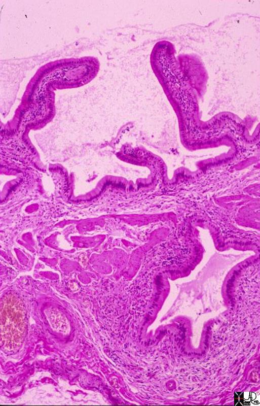
| This is a low power photomicrograph showing full thickness gallbladder wall. The lumen is the empty part with mucosal fronds projecting into it. The prominent hole deep in the wall is a Rokitansky-Aschoff sinus. This one is seen in cross section, but in vivo the sinuses are in continuity with the lumen because they are diverticula, or protrusions of mucosa out through the muscular layer, possibly in response to increased pressure from gallstones. Note that the sinus is lined by a single layer of columnar epithelium, just like the lumen itself. Because they are lumenal projections, these sinuses become filled with bile. Gallstones can become impacted in them. In acute cholecystitis, inflammation can spread directly through the wall following the Rokitansky-Aschoff sinuses. In chronic cholecystitis as seen here, the sinus is associated with some fibrosis and an infiltrate of mononuclear inflammatory cells.
11931.8s gallbladder chronic cholecystitis histopathology Courtesy Barbara Banner MD |
Generalized form
|
|
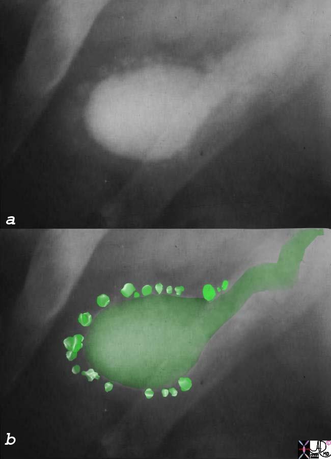
| Oral cholecystography is not used in contemporary imaging but it best demonstrates the diverticuli and the rosary resemblance. The diverticuli extend peripheral to the gallbladder lumen.
04743c01s gallbladder contracted outpouchings diverticula prominent Aschoff Rokitansky sinuses hyperplastic cholecystosis hyperplastic cholecystoses adenomyomatosis oral cholycysogram post fatty meal courtesy Ashley Davidoff MD copyright 2008 |
 Aschoff-Rokitansky Sinuses
|
Aschoff-Rokitansky Sinuses |
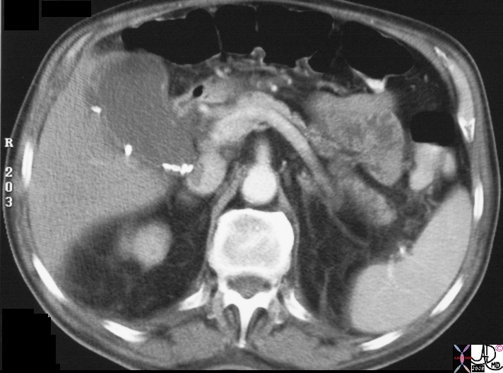
| Calcifications in non dependant position, peripheral to the lumen are characteristic of stones in rokitansky Aschoff sinuses are found in aassociation with calcified stones that are dependant in position inside the lumen and are characteristic of cholelithiasis.
16176bs gallbladder outpouchings diverticula prominent Aschoff Rokitansky sinuses thickened wall hyperplastic cholecystosis hyperplastic cholecystoses adenomyomatosis calcifications in wall courtesy Ashley Davidoff MD copyright 2008 |
|
Rokitansky-Aschoff Sinuses |
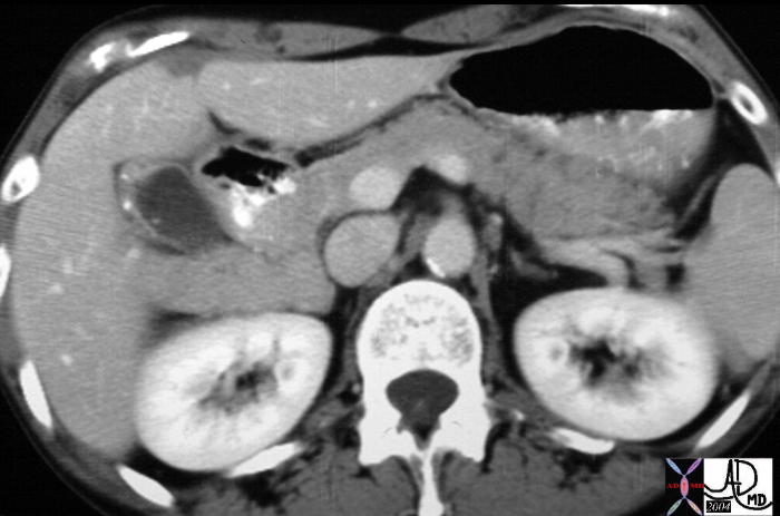
| CT scan of this 74 year old male shows large fluid filled diverticuli reminiscent of deep Rokitansky Aschoff sinuses.
16177s gallbladder outpouchings diverticula prominent Aschoff Rokitansky sinuses thickened wall hyperplastic cholecystosis hyperplastic cholecystoses adenomyomatosis calcifications in wall courtesy Ashley Davidoff MD copyright 2008 |
Localized form
Calcific Foci in the Wall with Focal Thickening |
| Calcifications peripheral to the lumen in the fundus and focal thickening are characteristic CT findings of adenomyomatosis.
19701 gallbladder + wall + fx calcification + cholecystoses + hyperplastic cholecystoses cholesterolosis imaging radiology CTscan Aschoff -Rokitansky sinuses hyperplastic cholecystosis hyperplastic cholecystoses adenomyomatosis cholesterolosis |
|
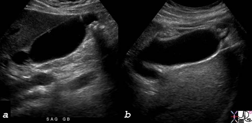
Rokitansky-Aschoff- Sinus filled with stones in the Fundus |
| A single fundal diverticulum is seen in this longitudinal ultrasound of the gallbladder. The diverticulum contains multiple shadowing stones.
78348c02s gallbladder outpouchings diverticula prominent Aschoff Rokitansky sinuses thickened wall hyperplastic cholecystosis hyperplastic cholecystoses adenomyomatosis calcifications in fundus courtesy Ashley Davidoff MD copyright 2008 |
|
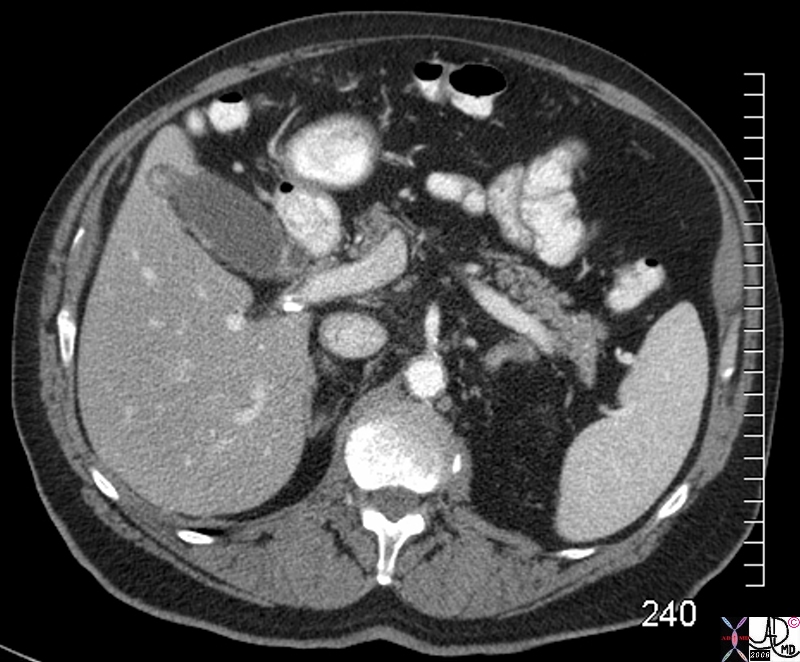
|
| A single fundal diverticulum is seen in this transverse section through the abdomen on an abdominal CTscan. The diverticulum either contains a low density calcification or has a hyperemic mucosa.
78356s gallbladder outpouchings diverticula prominent Aschoff Rokitansky sinus in fundus containing stones calcifications thickened wall hyperplastic cholecystosis hyperplastic cholecystoses adenomyomatosis calcifications in fundus CTscan courtesy Ashley Davidoff MD copyright 2008 |
 Focal Rokitansky-Aschoff Sinus in the Fundus