
Copyright 2008
Alok Anand Ashley Davidoff MD
Introduction
The character of the gallbladder offers clues to changes in the basic principles we have discussed. Several of the pathologic findings are mentioned below, though it is important to note what normal variation covers.
The bile is normally a lower density from the surrounding liver tissue, and is usually of a water density. Since it contains mostly water and some element of fat metabolism, it is generally relatively low and close to 1-5 HU.
The lumen should not have filling defects or focally enhancing areas. On ultrasound the luminal contents should be anechoic. The wall is typically thin (2-3mms), and blood flow is not normally exhibited to the wall.
Stones
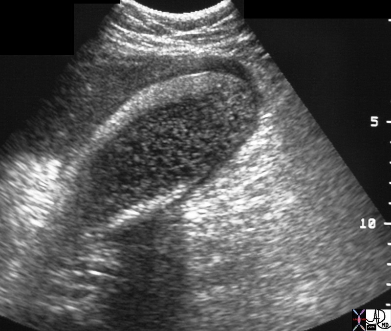
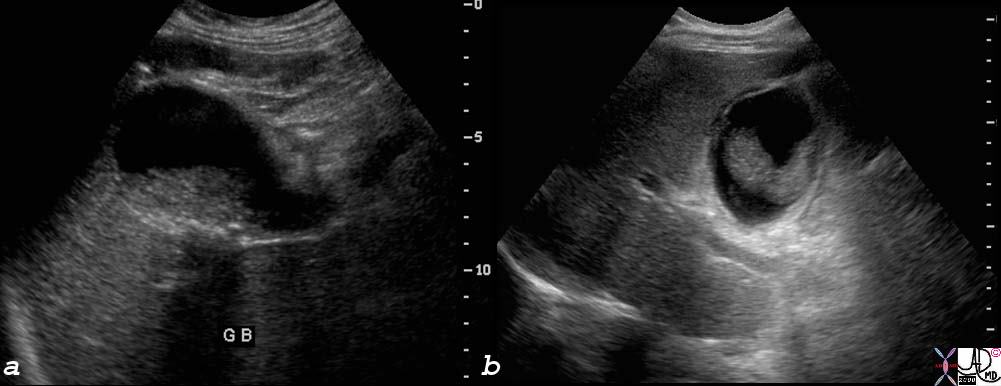
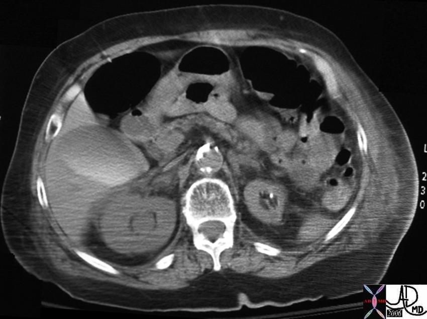
Obesity and Cholesterol Stone |
| 82205.8s 82206.8s 54 female mildly enlarged non tender gallbladder no abnormality seen on CTscan but large stone seen on USscan consistent with cholesterol stone isodense on CT shadowing on ultrasound atony obese Courtesy Ashley Davidoff MD Copyright 2008 |
Floating and Gas Filled |
| 26540 gallbladder + fx filling defects + fx air + fx floating + dx cholelithiasis + imaging radiology CTscan C- |
Air
Emphysematous Cholecystitis |
| 16195c02.8s elderly patient with heart disease and significant atherosclerotic disease gallbladder lumen is filled with air wall linear air collection bubbles of air air fluid level dx emphysematous cholecystitis CTscan Courtesy Ashley Davidoff MD copyright 2008 |
Emphysematous Cholecystitis |
| 61 year old who presents with abdominal pain, acute inferior myocardial infarction, with a background history of type II diabetes 31093 Courtesy Ashley Davidoff MD code pancreas fx induration dx acute pancreatitis code gall bladder wall fx air dx emphysematous cholecystitis size |
Emphysematous Cholecystitis |
| 61 year old who presents with abdominal pain, acute inferior myocardial infarction, with a background history of type II diabetes
31095c Courtesy Ashley Davidoff MD code pancreas fx induration dx acute pancreatitis code gall bladder wall fx air dx emphysematous cholecystitis size |
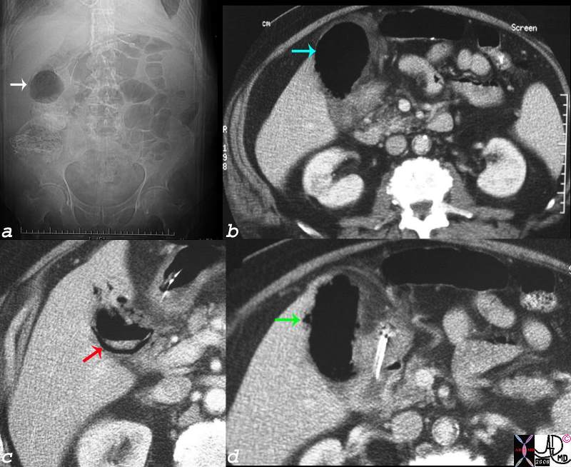
Gallstone Ileus |
| 31127c01.8s gallbladder stones air adherent to duodenum small bowel dilatation calcification stone gallstone ileus CTscan Courtesy Ashley DAvidoff copyright 2008 |
Fat
Crystalline Floaters |
| 77753c01.8s young female right upperquadrant tenderness RUQ gallbladder echogenic irregular fluid fluid layer conforming to the shape of the gallbladder wall thickened linear echoes floaters crystalline SG less than bile cholesterol crystals specific gravity forces space cholelithiasis stones small acute cholecystitis USscan ultrasound Courtesy Ashley Davidoff MD For Radiologists and Detectives |
Crystalline Floaters |
| 77753c02.8s young female right upperquadrant tenderness RUQ gallbladder echogenic irregular fluid fluid layer conforming to the shape of the gallbladder wall thickened linear echoes floaters crystalline SG less than bile cholesterol crystals specific gravity forces space cholelithiasis stones small acute cholecystitis USscan ultrasound Courtesy Ashley Davidoff MD For Radiologists and Detectives |
Tiny Calcified Floaters |
| 45092 45092b02 gallbladder fx cholelithiasiscompletely occupying gallbladder lumen CTscan Courtesy Ashley Davidoff MD |
Tiny Calcified Cholesterol Floaters |
| 25306.8s small cholesterol crystals stones floaters hyperemic wall chronic cholecytitis gas air cholelithiasis CTscan Courtesy Ashley Davidoff MD copyright 2008 |
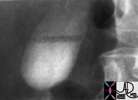
Stones Floating on Surface – Cholesterol Content – Historical Image |
| 04730 gallbladder floating small filling defect cholesterol stones cholelithiasis OCG oral cholecystogram imaging radiology historical |
Fluid
Hepatization
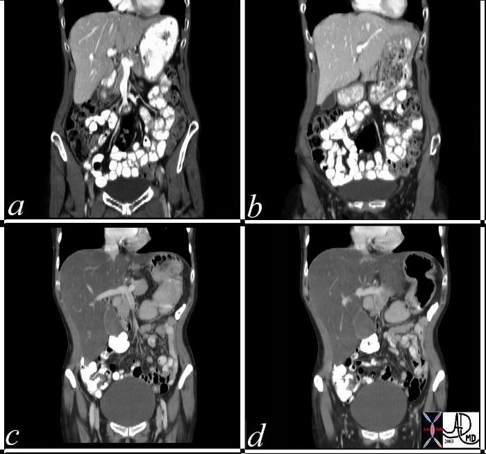
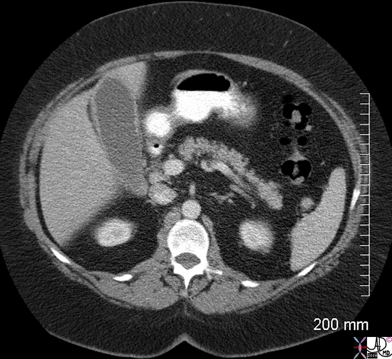
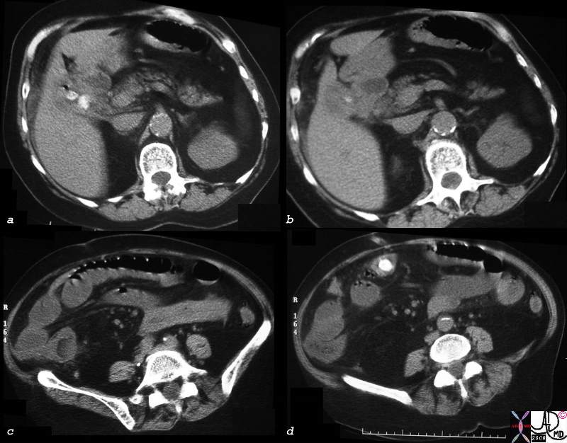
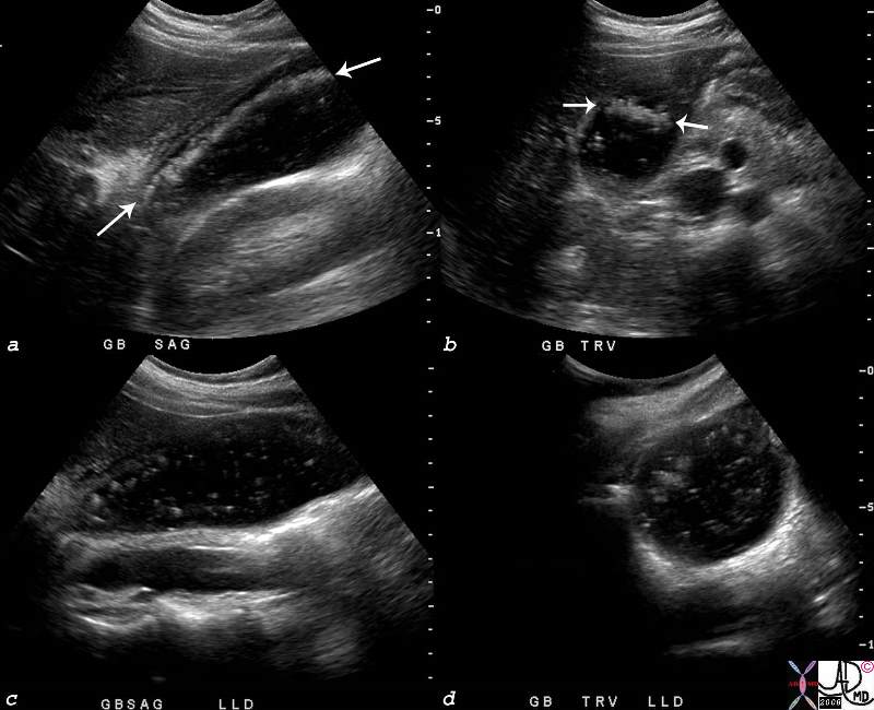
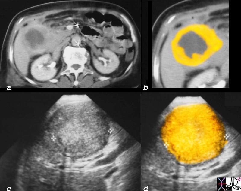
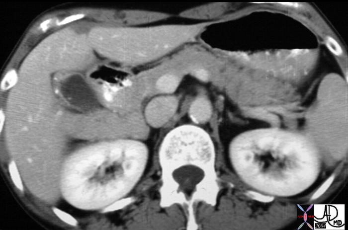
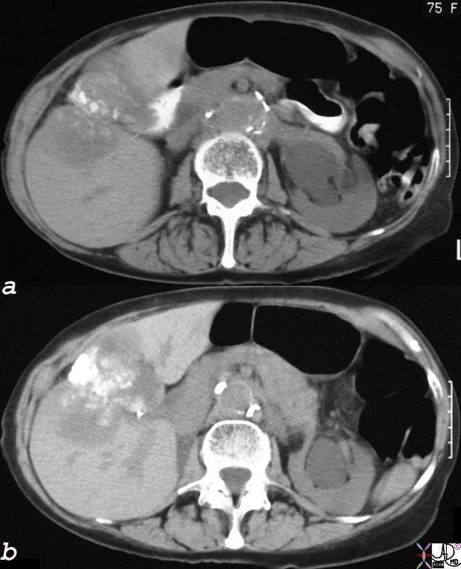
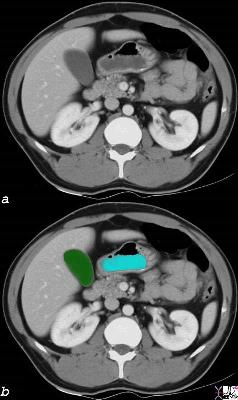
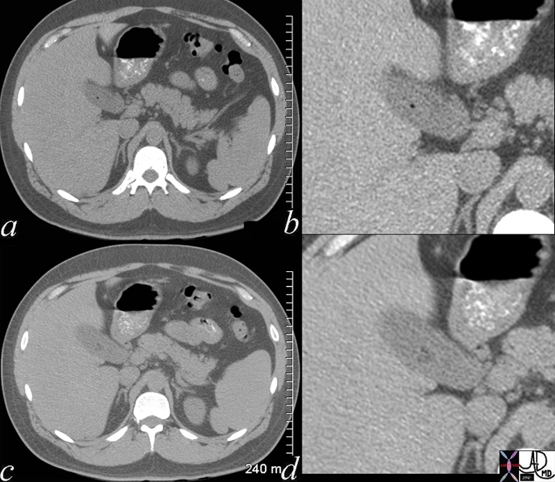
Hepatization of the Gallbladder Steatosis of the Liver |
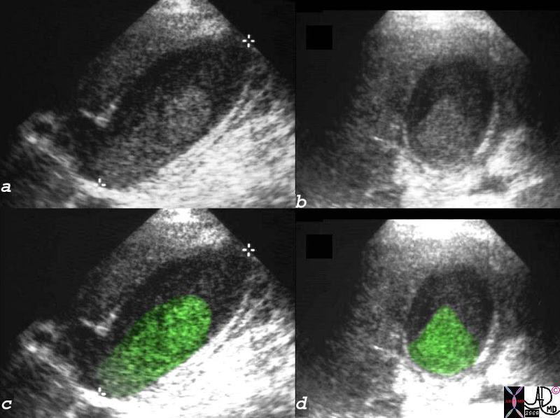
| Images a and b are from a normal patient. Note the density of the liver and compare to the density of the gallbladder in image b. The patient in c and d has diffuse steatosis and an enlarged liver. Note the difference in the liver density of the two cases and the difference in the relative densities of the gall bladders. The liver is normal in size in case A (a,b) and enlarged in case B (c.d) The patient in c and d is known to abuse both alcohol and drugs. Courtesy Ashley Davidoff MD 37833c code liver gallbladder steatosis fatty liver normal large |
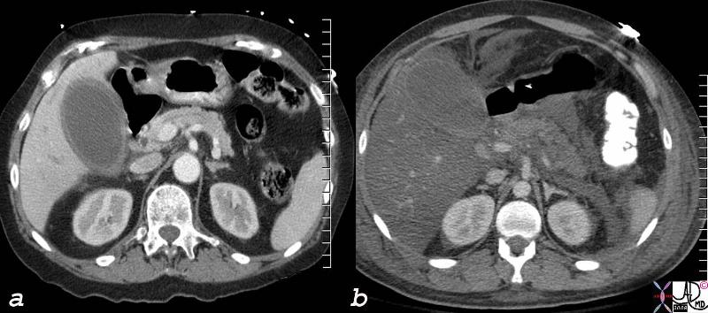
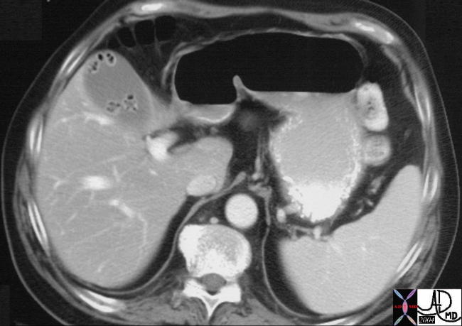
Hepatization of the Gallbladder Steatosis of the Liver |
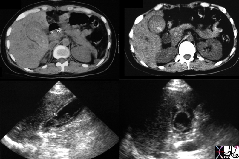
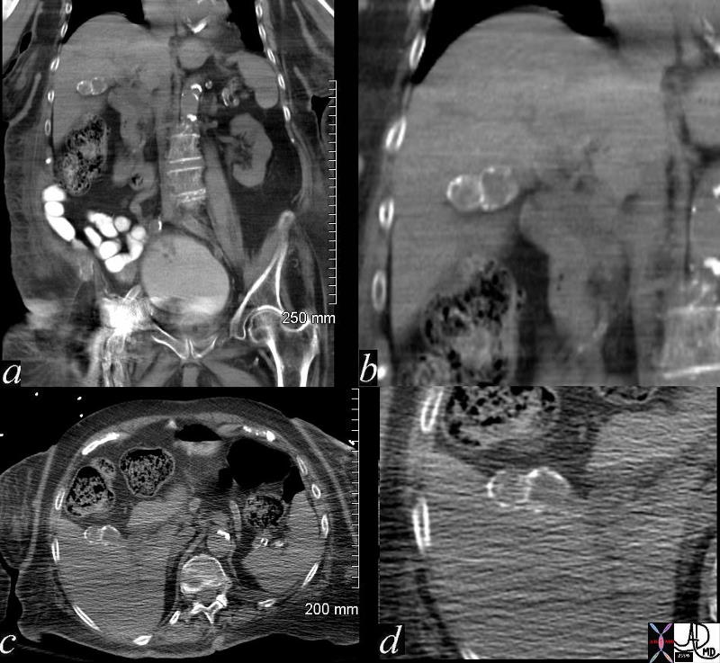
| 78606c.8s liver normal liver abnormal fatty change 777017 = a = acalculous cholecystitis b= pancreatitis with steatosis hepatization of the gallbladder density of the liver is the same as the bile in the gallbladder CTscan Courtesy Ashley Davidoff MD copyright 2008 |
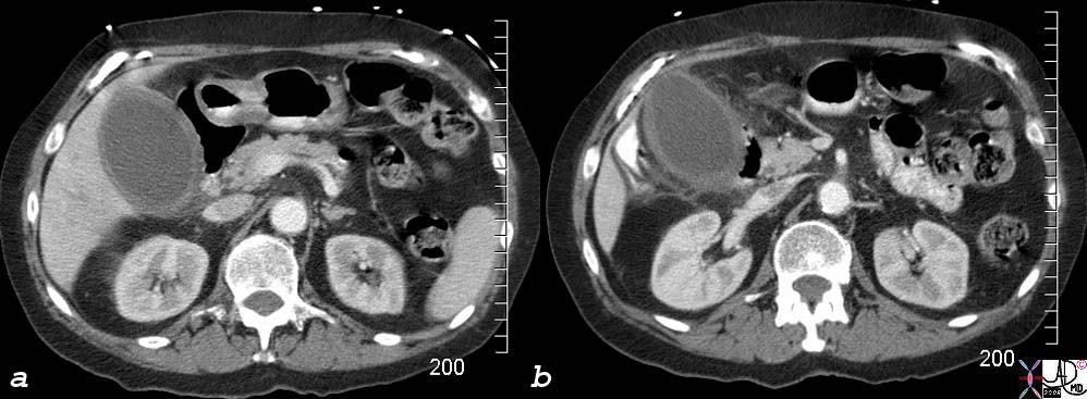
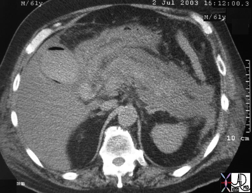
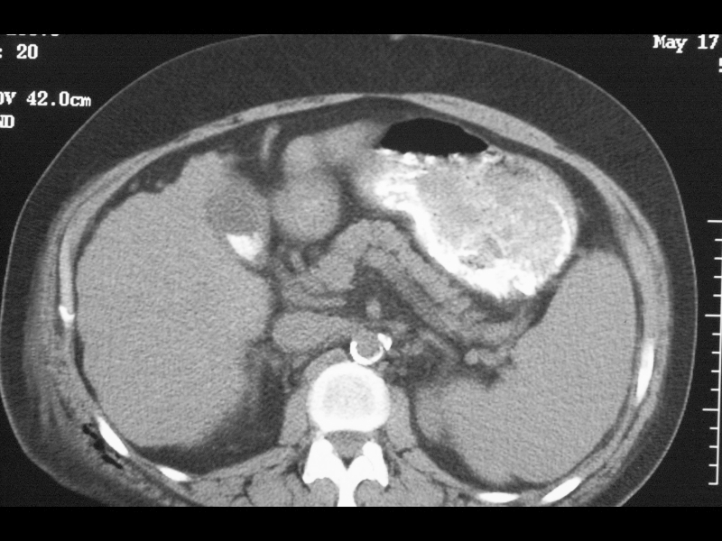
RUQ Pain, Distended Gallbladder, Indurated Fat |
| 77717c02s right upper quadrant pain ICU setting RUQ pain gallbladder distended thick walled hyperemic enhancing wall pericholecystic induration dirty fat stranding acalculous cholecystitis Courtesy Ashley DAvidoff MD copyright 2008 |
Semifluid
Empyema |
| 48010.800 gallbladder fx thick walled shadowing cholelithiasis complex fluid dx gallbladder empyema |
Sludge
Black – like Crank Case Oil |
| 26067 bile black crankcase oil dx acalculous cholecystitis body fluid Laboratory Test Courtesy Ashley DAvidoff MD |
Acaclculous Cholecystitis |
| 77680c01.8 right upper quadrant pain RUQ pain ICU setting gallbladder distended sludge cholestasis complex fluid in the gallbladder fossa acalculous cholecystitis USscan Courtesy Ashley Davidoff MD copyright 2008 |
Tumefactive Bile
Acaclculous Cholecystitis |
| 16136c03.8s gallbladder prolonged ICU stay right upper quadrant pain right upper quadrant tenderness cholestasis enlarged tumefactive bile sludge thick walled acalculous cholecystitis percutaneous cholecystostomy ultrasound USscan Courtesy Ashley Davidoff MD copyright 2008 |
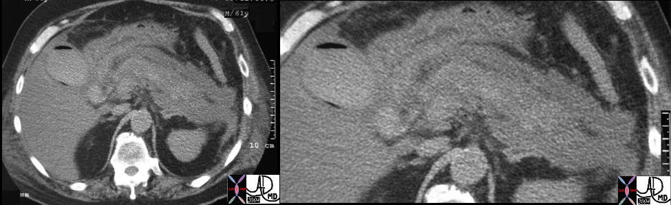
RUQ Pain, Distended Gallbladder, Tumefactive Bile – same patient |
| 77717c01s right upper quadrant pain ICU setting RUQ pain gallbladder distended thick walled sludge cholestasis tumefactive bile fluid in gallbladder fossa small shadowing stones acalculous cholecystitis shape of tumefactive bile in the gallbladder has fetus like formation USscan ultrasound Courtesy Ashley DAvidoff MD copyright 2008 |
Soft Tissue
Carcinoma – Local Invasion and Separate Liver Mestastasis |
| 17280c02b01.8s gallbladder liver mass local invasion cholelithiasis metastasis carcinoma primary gallbladder gallbladder fossa Courtesy Ashley Davidoff MD copyright 2008 |
Lumenal Invasion |
| 16254c04.8s gallbladder space occupation gallbladder carcinoma by CT it appears as a low density centrally and enhancing soft tissue peripherally by USscan looks like the whole lumen is filled with soft tissue tumor question of delayed tumor enhancement vs necrosis CTscan USscan ultrasound Courtesy Ashley Davidoff copyright 2008 |
Hyperdense
Stones
|
Aschoff-Rokitansky Sinuses |
| 16176bs gallbladder outpouchings diverticula prominent Aschoff Rokitansky sinuses thickened wall hyperplastic cholecystosis hyperplastic cholecystoses adenomyomatosis calcifications in wall courtesy Ashley Davidoff MD copyright 2008 |
Layering Hyperdense |
| 78352.8s obese female gallbladder layering hyperdense stones cholelithiasis hyperemic wall hyperemic mucosa and submucosa chronic cholecystitis CTscan Courtesy Ashley Davidoff MD copyright 2008 |
Calcific Foci in the Wall with Focal Thickening |
| 19701 gallbladder + wall + fx calcification + cholecystoses + hyperplastic cholecystoses cholesterolosis imaging radiology CTscan Aschoff -Rokitansky sinuses hyperplastic cholecystosis hyperplastic cholecystoses adenomyomatosis cholesterolosis |
Echogenic Foci in the Wall with Ringdown Artifact |
| 82101cs 34 female gallbladder adenomyomatosis ring down artifact hyperplastic cholecystoses calcifications stones Aschoff-Rokitansky sinuses USscan ultrasound Courtesy Ashley DAvidoff MD copyright 2008 |
|
Porcelain Gallbladder |
| 47683c01 gallbladder fx calcification in wall calcified wall dx porcelain gallbladder CTscan Davidoff MD |

Milk of Calcium Bile |
| 16489 gallbladder + milk of calcium bile +liver micronodular cirrhosis imaging radiology CTscan |
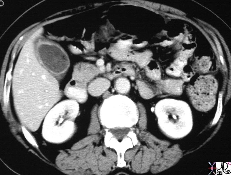
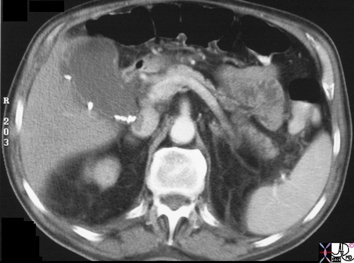
Renal Failure |
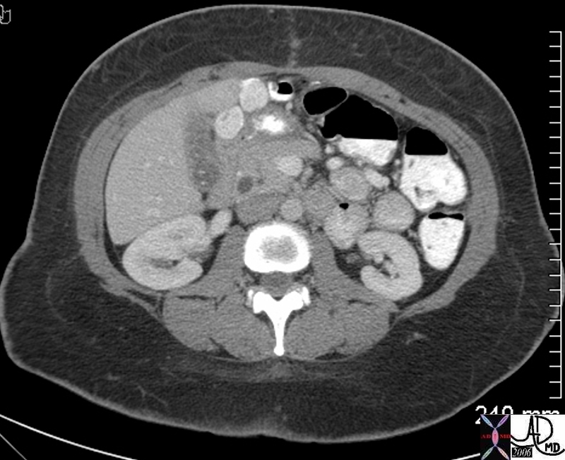
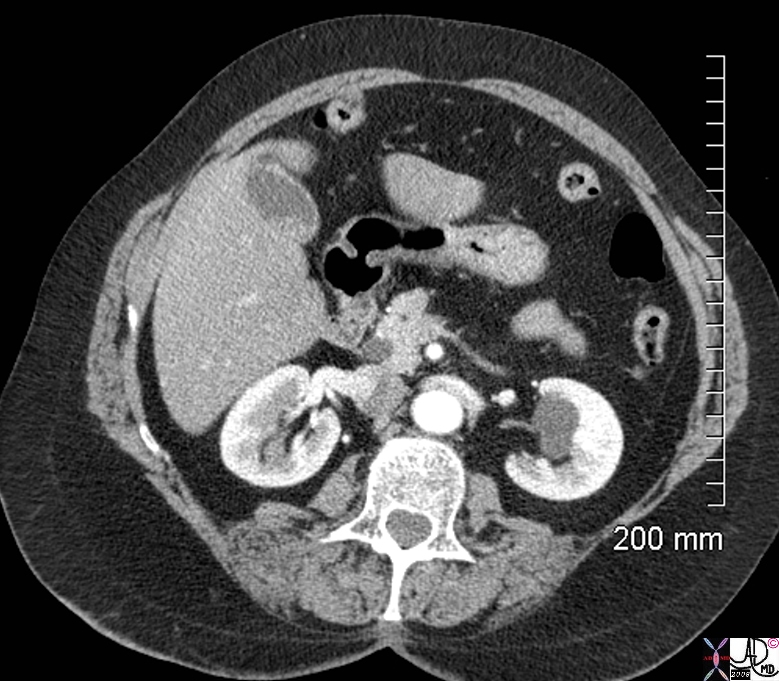
| 24075 81Y F with LLQ pain s/p cardiac catheterization gallbladder fx fluid -fluid level fx high density sediment fx vicarious excretion of contrast s/p heart catheter kidney fx small left kidney fx enlarged right kidney with hydronephrosis muscle fx hematoma dx pelvic hematoma CTscan Courtesy Ashley Davidoff MD |
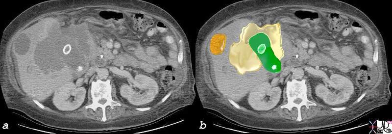
Dystrophic Calcification – Carcinoma of the Gallbladder Extending into the Gallbladder Fossa |
| 24404c.8s 75 female gallbladder calcification adjacent mass in the liver local invasion into the gallbladder fossa dystrophic calcification probably mucinous adenocarcinoma of the gallbladder carcinoma stones cholelithiasis hydronephrosis |
Acute Hemorrhagic Cholecystitis |
| 81902s.8b01 elderly man with acuteright upper quadrant pain gallbladder wall mildly distended gallbladder hyperdense irregular wall dx acute hemorrhagic cholecytitis acute on chronic cholecystitis kidney cyst complex calcification grade 3 Bosniak CTscan Courtesy Ashley Davidoff MD copyright 2008 |
Hemorrhagic Cholecystitis |
| 16214c.8s gallbladder distended enlarged containing hyperdense material sludge differential diagnosis tumefactive bile cholestasis dx acute hemorrhagic cholecystitis CT scan USscan Courtesy Ashley Davidoff MD Copyright 200 |
Multiple small shadowing forming fluid fluid layer
Moderate size stones
sludge echogenic sediment/ bile sludge level / tumefactive/completely filling the gb/ mobility
Other fluids that look like sludge sand (non homogeneous and sghadows) /beam width artifact/ empyema/hemorrhage
polypoid lesions hemisherical highly reflective few mms in size no shadow
Ct – sludge has a slightly higer density than bile
Normal Fasting Gallbladder |
| 18138c.8s gallbladder fasting normal size about 100ccs fluid secretions in the stomach anatomy size physiology function shape position character water density CTscan Courtesy Ashley Davidoff MD copyright 2008 |
Clear Fluid |
| 47018 gallbladder normal shape pear shaped anatomy nrmal USscan Davidoff MD |
SG less than ERCP Contrast |
| 82140.8s patient s/p ERCP gallbladder patient supine contrast layer posterior sediment bile supernatant SG specific gravityis of bile is less thanthat of contrast forces Courtesy Ashley DAvidoff MD copyright 2008 |
Applied Biology
Lumen
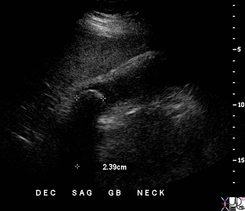
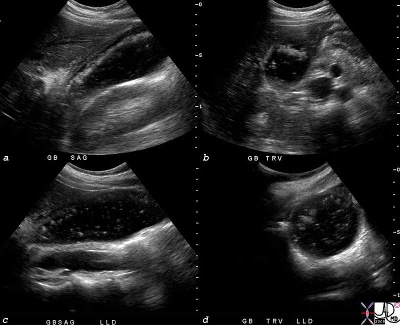
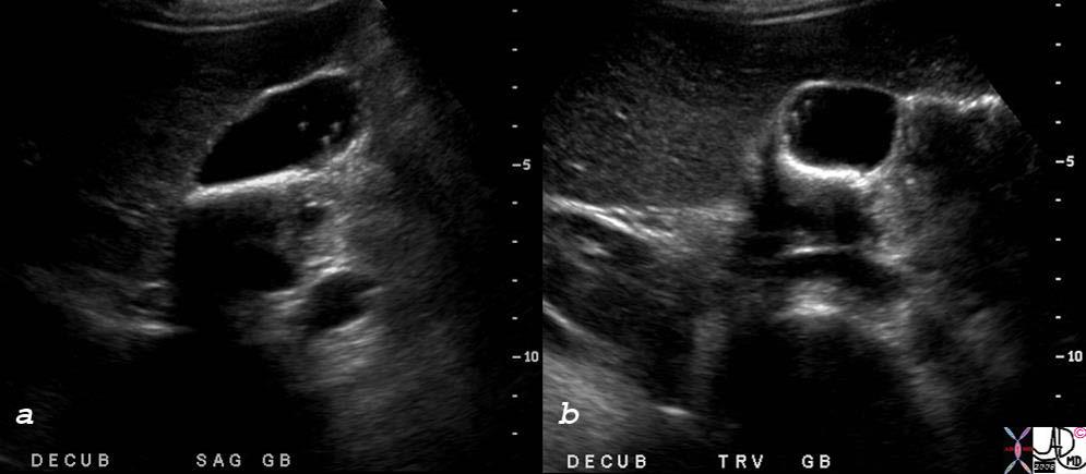
Sludge – Almost Fluid |
| 78365c.8s gallbladder sludge cholestasis fluid fluid level USscan ultrasound Courtesy Ashley Davidoff MD copyright 2008 |
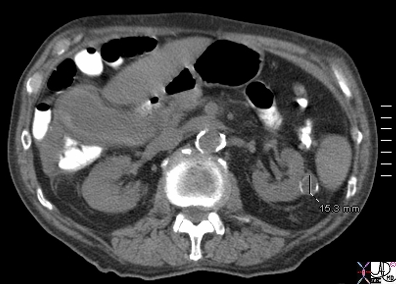
Subtle Character Changes |
| 70269c02 gallbladder air methane nitrogen gas cholesterol stone isodense with bile fat subtle change CTscan |
|
Dilated in Acalculous Cholecytitis and Dilated in Fatty Liver |
| 78606c.8s liver normal liver abnormal fatty change 777017 = a = acalculous cholecystitis b= pancreatitis with steatosis hepatization of the gallbladder density of the liver is the same as the bile in the gallbladder CTscan Courtesy Ashley Davidoff MD copyright 2008 |
|
Empyema |
| 48010.800 gallbladder fx thick walled shadowing cholelithiasis complex fluid dx gallbladder empyema |
Wall
Porcelain Gallbladder |
| 47683c01 gallbladder fx calcification in wall calcified wall dx porcelain gallbladder CTscan Davidoff MD |
|
Milk of Calcium Bile |
| 16489 gallbladder + milk of calcium bile +liver micronodular cirrhosis imaging radiology CTscan |

Post Op Bed – Gelfoam |
| 46625 gallbladder fossa s/p cholecystectomy with gelfoam packing of fossa fx echgenic collection in gallbladder fossa dx air within gelfoam USscan Davidoff MD |
References
Catalano et al MR Imaging of the Gallbladder RadioGraphics 2008;28:135-155
On T1-weighted images, the gallbladder wall has intermediate signal intensity and enhances uniformly after the administration of gadolinium-based contrast material. The portion of the gallbladder wall adjacent to the liver may not be well appreciated owing to similar enhancement of the wall and the liver parenchyma. On T2-weighted images, the gallbladder wall has low signal intensity and stands out against the bright visceral fat. The wall adjacent to the liver cannot be identified as a separate structure.
Normal bile appears uniformly bright with T2-weighted sequences. On T1-weighted images, bile varies greatly in signal intensitydepending on its concentration.
During fasting, bile undergoes a process of concentration. Water is reabsorbed and the concentration of cholesterol and bile salts increases, leading to a shortened T1 relaxation time and, consequently, to bright bile on T1-weighted images. A layering effect is sometimes observed, with concentrated and denser bile in the dependent position.
Gallbladder sludge may have similar signal intensity characteristics, namely, iso- to mild hyperintensity on T2-weighted images andhyperintensity on T1-weighted images
|
T1 |
T2 |
||
| Wall | pre Gad | intermediate | low
wall on liver side may not be appreciated |
| post Gad | uniform enhancement
wall on liver side may not be appreciated |
||
| Lumen (bile) | non fasting | variable depending on concentration | bright |
| fasting | bright, sometimes layering effect is seen | bright | |
| sludge | hyperintensity with layering | iso to mild hyperintensity | |
References
Hsu-Chong et al Floating Gallstones in Bile without Added Contrast AJR 1986
Tera H. Stratification of Human Gallbladder Bile In Vivo Acta Chirg Scan (Suppl) 1960 256: 9-85 (not available Pubmed)
Hawk Practical Physiological Chemistry By Philip Bovier Hawk, Olaf Bergeim Published by Blakiston, 1918