
The Common Vein Copyright 2008
Alok Anand
Definition
Cholelithiasis or gallstones are the result of a metabolic disorder of bile metabolism caused by an increase in the insoluble portions of bile relative to the solutes which keep them in solution, and resulting in precipitated solutes of biliary secretions in the gallbladder. Gallstones are more common in women, and are associated with diseases relating to cholesterol metabolism, diabetes, mellitus., heolytic anemai, cirrhosis.
The resulting concretions are common in the Western world, (8% in the population at large and 10-15% in ages 40-60)) and more commonly than not of no clincial significance. However occasionally they may impact in the neck, cystic duct or common bile duct, causing obstruction in the biliary tract. The obstruction progresses to inflammation and acute cholecystitis, or cholangitis may result.
There are three forms of stones:
Cholesterol
Pigment
Mixed
The most common stones are cholesterol stones, which account for 75-80% of cases. The next are pigment stones, which account for approximately 15% of all cases. The remainder of cases are mixed stones, representing a combination of the previous two. An aberration of cholesterol metabolism causes an imbalance between cholesterol and the bile acids, phospholipids and lecithin which keeps the cholesterol in solution. Similarly, increased bilirubin metabolism from the breakdown of hemoglobin, usually due to hemolysis, causes in increased excretion of bile pigments, leading to stone precipitation. These are called black pigment stones. An additional type of pigment stone is the brown pigment stone. This is associated with intra-hepatic infection, and is seen mostly in Asian populations.
Structurally, cholelithiasis is characterized by the presence of rock-like crystals in the dependent portion of the gallbladder. There is no characteristic change in the size or shape of the gallbladder unless an obstruction occurs. The stones vary from small crystals in the submms range to very large stones which may occupy the whole gallbladder. They be round, oval, or faceted. Calcification may be dense, or minimal, diffuse, laminated, or heterogeneous
Although there is no functional change with the presence of stones in the gallbladder itself, in later phases obstruction can impair excretion of bile salts and digestion. Aberrations in metabolism of cholesterol and hemoglobin predispose to the formation of gallstones.
Clinical presentation of patients varies with the staging of disease:
Asymptomatic: stones are merely present in the gallbladder and may be incidental findings on abdominal imaging.
Symptomatic: patients may have colicky RUQ pain, often radiating to the right shoulder (transient impaction without inflammation)
Complicated: RUQ pain, fever/leukocytosis (impaction with inflammation)
The symptomatic stage is characterized by 1-5 hour bouts of pain, usually after meals, and may recur. It may progress to complicated stage, where pain is of longer duration and accompanied by symptoms of infection.
Diagnosis is easily detected by ultrasound, (95%), but are seen by CT as well. (80-85%),. The stomes that are not seen by CT are usually cholesterol stones which are isodense with bile and very small non calcified stones. In the absence of obstruction, scintillography with HIDA scan provides little information, though in patients who have progressed to symptomatic and complicated stages, it is the most specific study for the diagnosis of an impacted stone in the neck or cystic duct.
Treatment depends on the staging. If asymptomatic and found incidentally, patients may be followed. If symptomatic, patients may have elective cholecystectectomy. In the complicated stage, patients are hospitalized, with IV antibiotics. Elective cholecystectomy is perfgormed after a “cool-down” period.
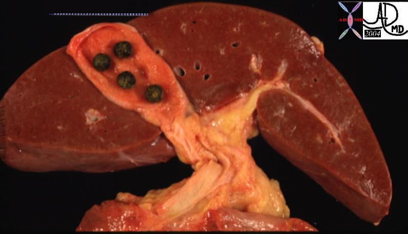
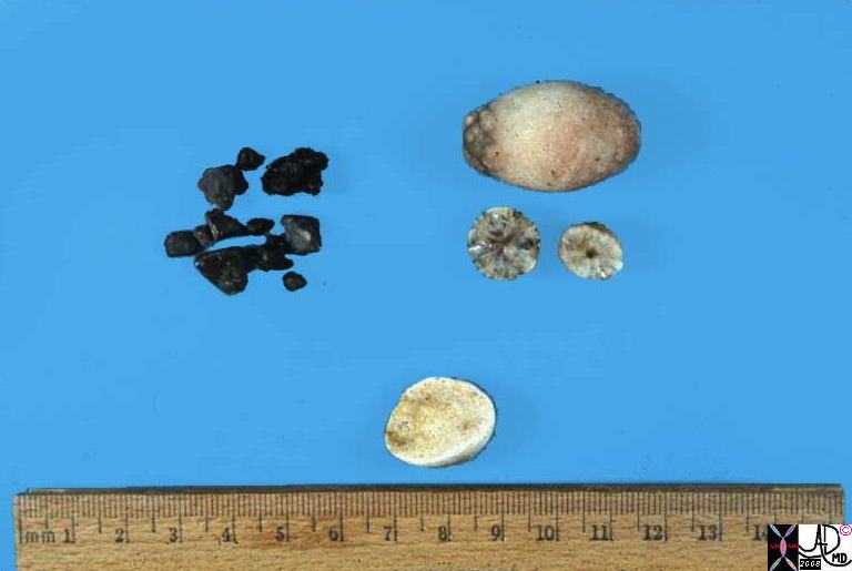 Types of Stones Types of Stones |
| This photograph shows examples of the three types of pure gallstones. The multiple small green-black stones are pure bilirubinate – “pigment stones”. The egg-shaped stone and two smaller halves on cut surface are pure cholesterol stones. These are clear and crystalline, like quartz. The single round, opaque chalky-tan stone is pure calcium carbonate, like limestone. Gallstones of pure composition occur about 10% of the time.
gallbladder dx cholelithiasis grosspathology Courtesy Barbara Banner MD 11918.83s |
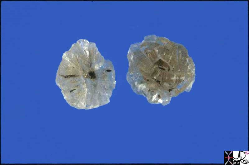 Cholesterol Stones Cholesterol Stones |
| This is a photograph of a pure cholesterol gallstone cut in half to show its crystalline beauty.
gallbladder dx cholelithiasis grosspathology Courtesy Barbara Banner MD 11919.81s |
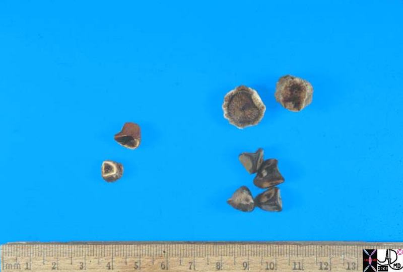 Types of Stones Types of Stones |
| This photograph shows examples of the most common gallstones, which are a mixture of bilirubin, cholesterol, calcium carbonate and other components. They are usually multiple, multifaceted, and about 0.5 –1.0 cm on a side. Note the small stone cut across which has a yellow rim and a dark green center. This is a combined stone with a bilirubinate center and a cholesterol-containing rim.
11920b03s gallbladder dx cholelithiasis grosspathology Courtesy Barbara Banner MD |
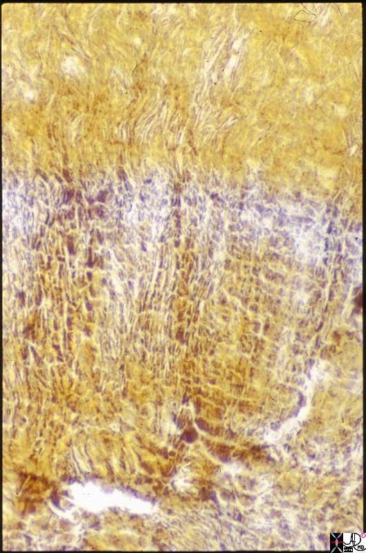 Micrsoscopic Appearance of a Stone Micrsoscopic Appearance of a Stone |
| This is a medium –power photomicrograph of a tiny gallstone which was present in a gallbladder and was included in the microscopic section of that gallbladder. The yellow color is the natural color of the stone, and there appears to be some linear or crystalline structure. Only the gallstone is present in the picture, no gallbladder tissue can be seen.
11921s gallbladder dx cholelithiasis histology Courtesy Barbara Banner MD |
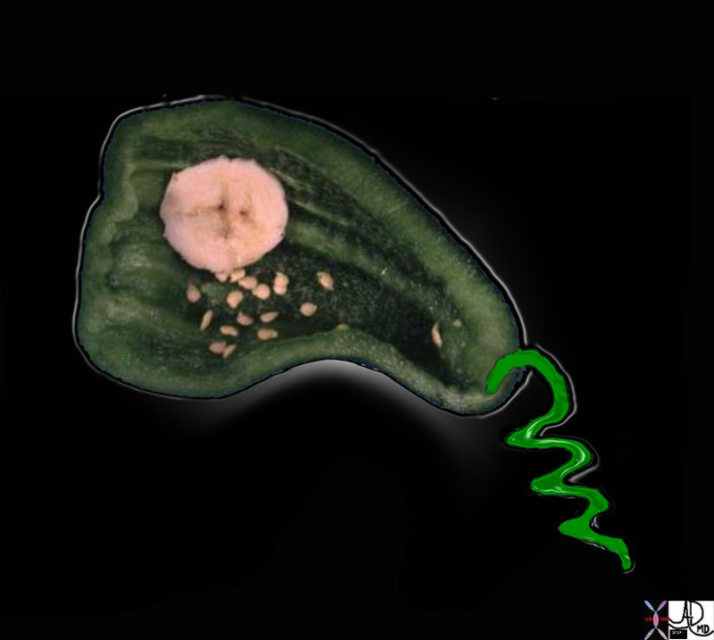
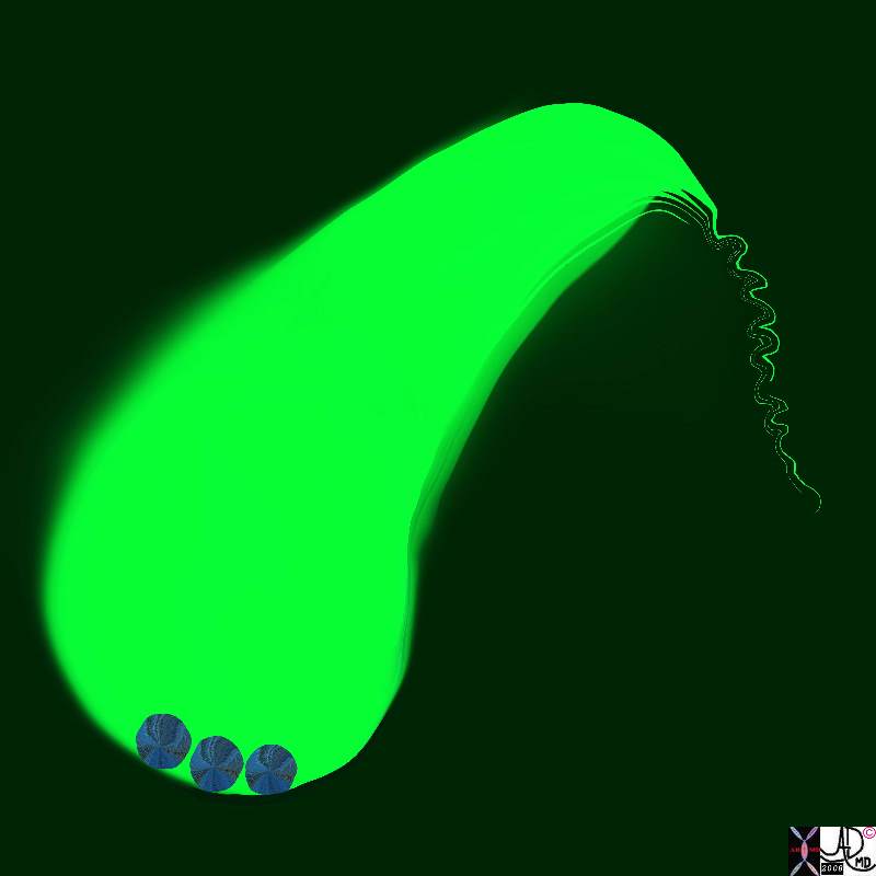
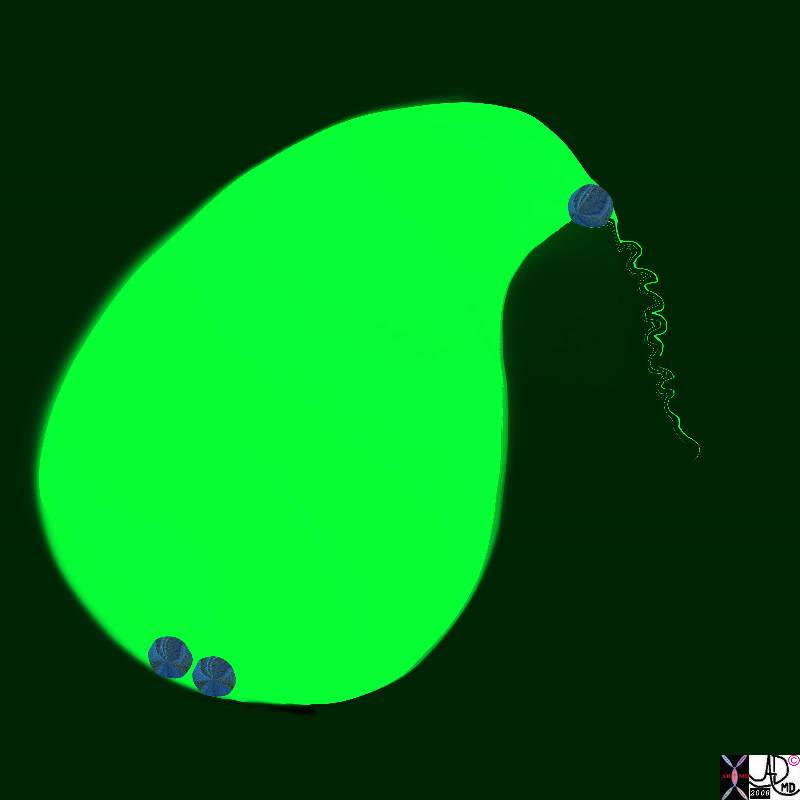
Greenpepper Gallbladder and Stones |
| 00404b01.800 gallbladder gallstones cystic duct cholelithiasis greenpepper banana Davidoff Art Davidoff MD |
Imaging Stones
Size
Small Floaters
Cholesterol Floaters |
| The smallest gallstones are seen as submm floating echogenic foci that represent crytals of cholesterol. They show comet tail artifact, and in this case occupy the entire lumen of the gallbladder.
Cholesterol floaters76356 gallbladder stones floaters crystaline tiny cholelithiasis ring down artifact floating USscan ultrasound Courtesy Ashley Davidoff MD copyright 2008 |
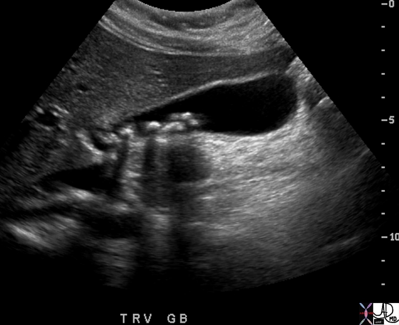
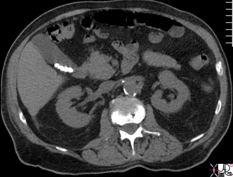
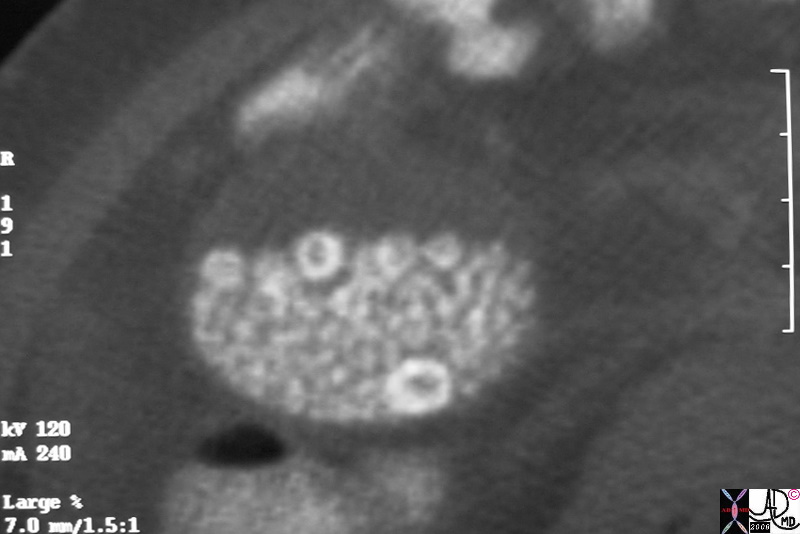
Small Sinkers
Small Calcified Stones |
|
Small calcified stones are more common than floaters, and in this case can be seen lying dependantly in the infundibulum of the gallbladder but at least three other smaller stones and two larger are seen in the region of the neck and cystic duct. 41164 Courtesy Ashley Davidoff MD code pancreas pancreatic calcifications code gallbladder calcified stones dx cholelithiasis dx chronic pancreatitis imaging radiology CTscan chronic inflammation metabolic |
Ill Defined Small Floaters
Tiny Calcified Cholesterol Floaters |
| 25306.8s small cholesterol crystals stones floaters hyperemic wall chronic cholecytitis gas air cholelithiasis CTscan Courtesy Ashley Davidoff MD copyright 2008 |
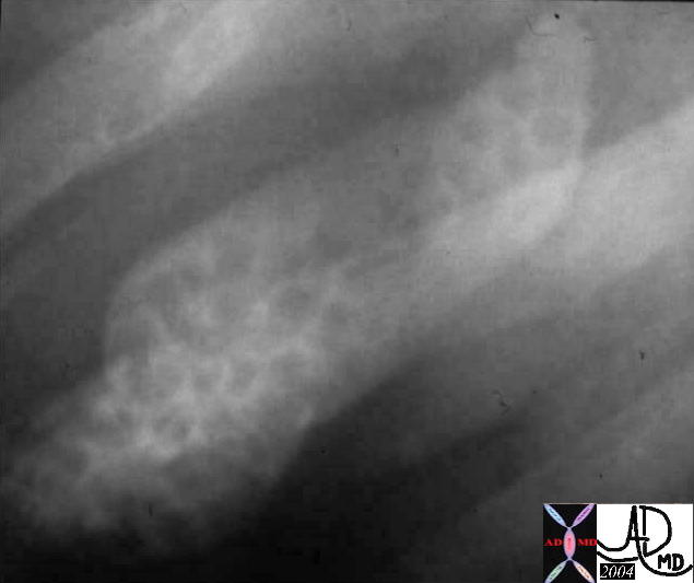
Well Defined Small Floaters
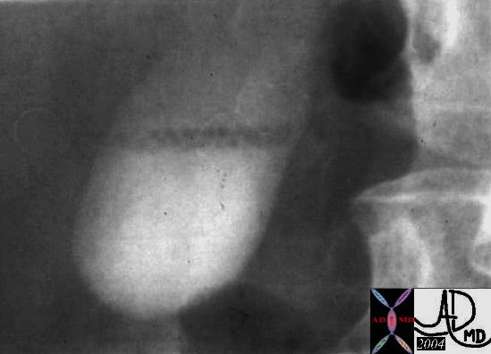
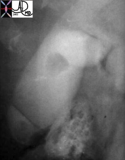
Stones Floating on Surface – Cholesterol Content – Historical Image |
|
04730 gallbladder floating small filling defect cholesterol stones cholelithiasis OCG oral cholecystogram imaging radiology historical |
Small Floaters That may Not Be Small Floaters
Tip of the Iceberg Stones |
|
In the CTscan there is a suggestion of tiny calcified floaters but this may also be deceiving since the calcifications may be flecks within larger isodense cholsterol stones. The latter appears to be the case since at the base of the gallbladder there is a rounded hypodense structure that is reminiscent of a cholesterol stone. 45092 45092b02 gallbladder fx cholelithiasiscompletely occupying gallbladder lumen CTscan Courtesy Ashley Davidoff MD |
Moderate Size Gall Stones
Position of the Gallbladder – Between Right and Left Lobes of the Liver |
| 13456 liver normal anatomy gallbladder stones cholelithiasis grosspathology |
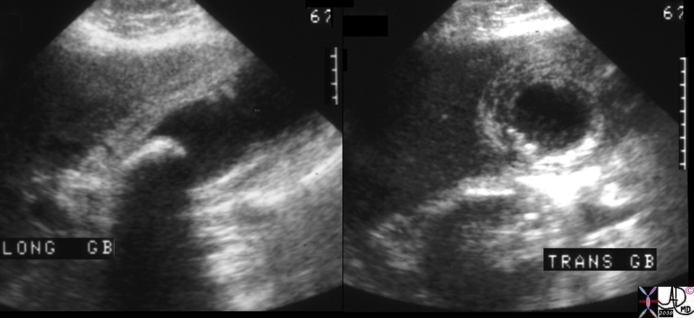
Multiple Medium Sized Shadowing Stones |
| 70026 gallbladder stones shadowing forces reflection transmission dx cholelithiasis USscan Davidoff MD |
Multiple Small to medium Stones Filling the GAllbladder Oral Cholecystogram – Historical Image |
| 04672 gallbladder multiple small stones cholelithiasis OCG oral cholecystogram imaging radiology |
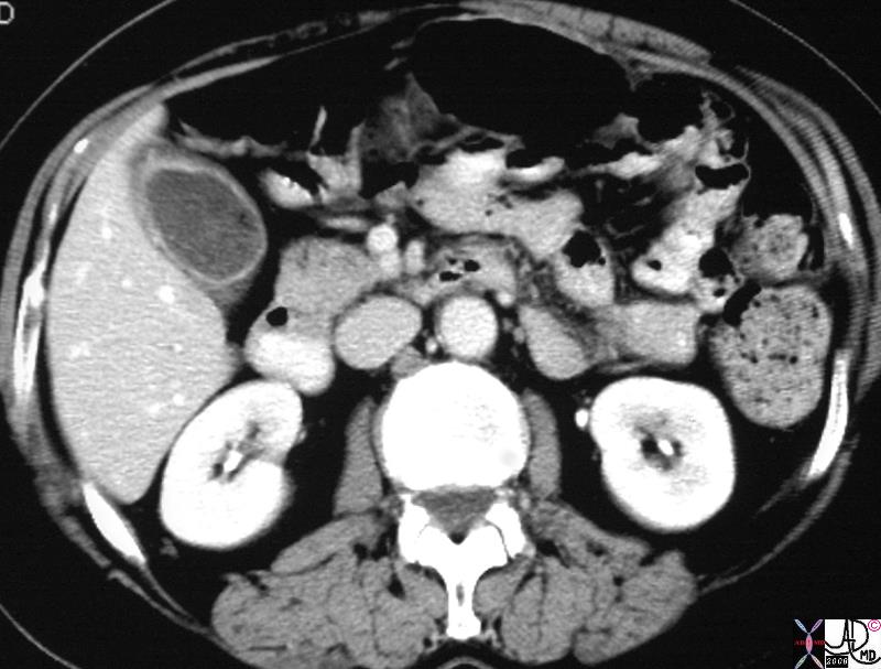
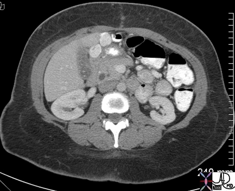
Small to Medium Sized Mobile Calcified Stones |
| 82167.8s gallbladder multiple stones cholelithiaisis dependant mobile CTscan Courtesy Ashley DAvidoff MD copyright 2008 |
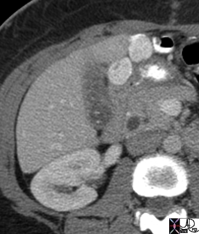
Single Moderate Size Enlarged Gallbladder with Phrygian Cap – Cholestasis |
| An intermediate size calcified stone in the 6-8mms range is seen lying dependantly in the large cholestatic gallbladder with a phrygian cap.
18261.8s gallbladder shape phrygian cap distended enlarged courtesy Ashley Davidoff MD copyright 2008 |
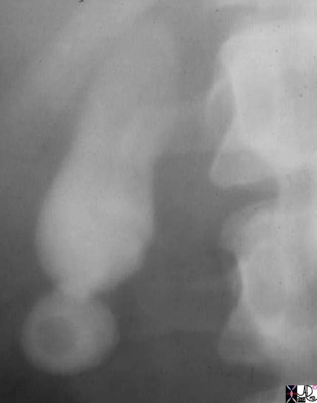
Moderaste Size Stone – Oral Cholecystogram – Historical Image
|
|
04407 gallbladder filling defect stones cholelithiasis OCG oral cholecystogram imaging radiology historical
|
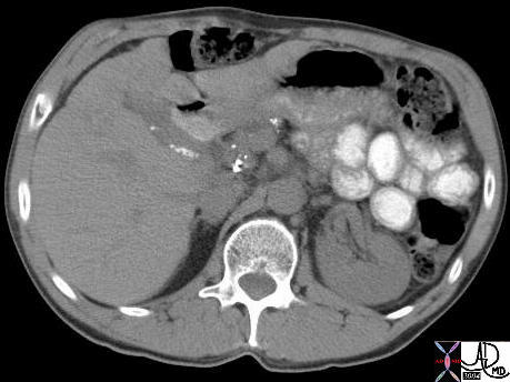
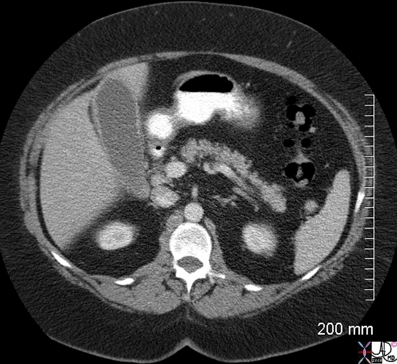
Large Stones
Large stones can occupy the entire gallbladder lumen and in general are less likely to cause obstruction of the neck. In general the larger the stone the less likely it will result in other significant disease. Large stones can uncommonly erode the wall and evolve into a gallstone ileus, but this is a rare entity.
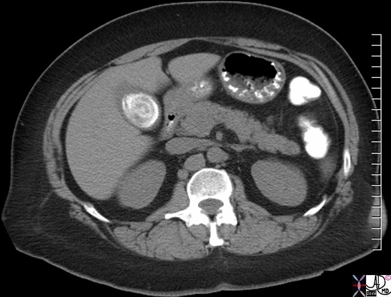
Large Lamellated Gallstone |
| The CT scan shows a large calcified layered stone occupying almost the entire lumen of the gallbladder.
abo82151.8s 51f obesea gallbladder stone large calcified lamellated cholelithiasis CTscan Courtesy Ashley DAvidoff MD copyright 2008 |
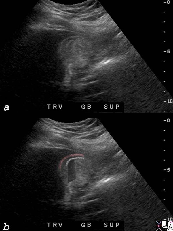
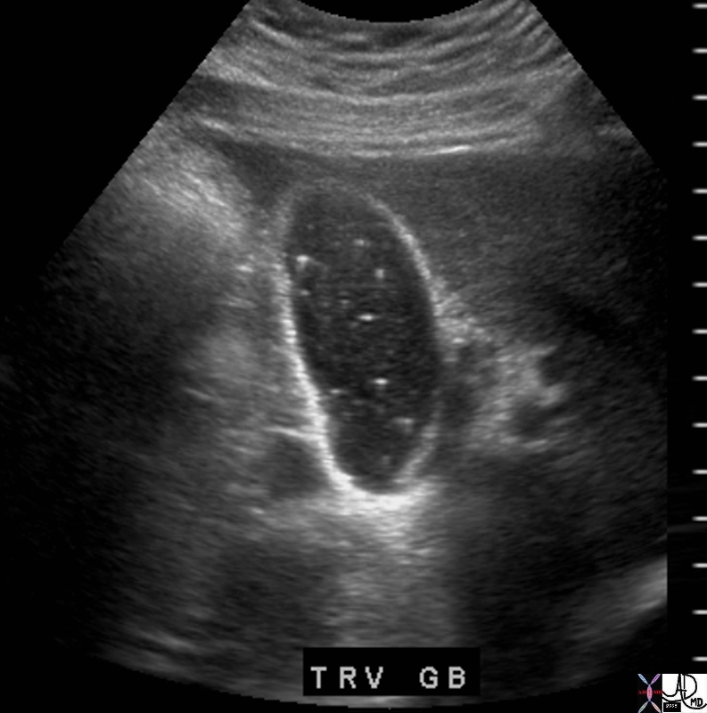
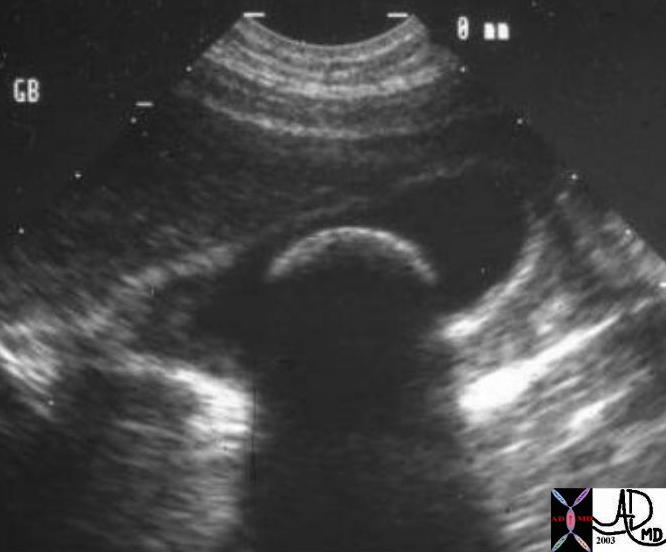
WES sign |
|
The ultrasound depicted above shows a large stone filling almost the entire gallbladder lumen so that the gallbladder is barely recognized. Familiarity with the WES sign allows recognition. The front wall of the gallbladder creates an echo (“W”) seen as a red overlay in b, the echo of the surface of the stone (“E”) seen as a white line in b is the second component, and then a shadow (“S”) is created by the stone. 82166c01s 27F gallbladder stone cholelithiasis WES sign wall = pink line echo = white line = front anterior echo of the stone black shadow = shadow of the large stone shadow USscan ultrasound Courtesy Ashley Davidoff MD copyright 2008 Courtesy Ashley Davidoff MD Copyright 2008 |
WES sign |
| 04733 gallbladder filling defects stones cholelithiasis WES sign USscan imaging radiology |
Applied Biology
Pregnancy and Cholelithisis |
| Gallbladder disese is common in pregancy probably due to stasis and smooth muscle relaxation due to changes in progesterone. The decreased emptying and decreased muscle tone leads to stasis. Sludge in pregnancy is therfore not uncommon. The estrogen and and progesterone cause an increased saturation of bile with cholestsrol making it lithogenic. In this case the cholesterol floaters are seen in the gallbladder which lay next to a the fetal abdomen. A cross section of the abdomen is noted and the fetus is compressing the the fundus of the gallbladder. The bile may return to normal in the postpartum state.
77625.8s gallbladder stones cholelithiasis cholesterol floaters pregnancy fetus gravid uterus deforming gallbladder USscan Ultrasound Courtesy Ashley Davidoff MD copyright 2008 |
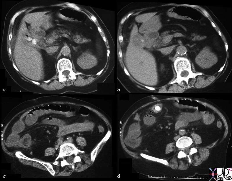
Small or Not So Small – Subtle Character Changes |
| A tiny air density in image a and b might suggest a small stone but it also could represent the tip of the iceberg of a larger isodense cholesterol stone. A subtle larger stone is seen in c and d that by virtue of its density confirms its cholesterol nature and larger size.
70269c02 gallbladder air methane nitrogen gas cholesterol stone isodense with bile fat subtle change CTscan |
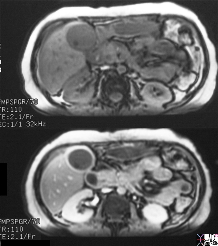
Small Stones Big Problems – Acute Cholecystitis |
| 16141c01s.8 gallbladder stones cholelithiasis hyperemic wall edematous wall small filling defects in dependant position in the infundibulum distended gall bladder edema in the wall fluid in the gallbladder fossa gbf normal bile duct MRI T1 weighted image with gadolinium and fat saturation T2 weighted MRCP normal pancreatic duct Courtesy Ashley Davidoff MD copyright 2008 |
|
|
| 78305.8s gallstone large stone cholelithiasis impacted in the infundibulum does not move with decubitus fluid in the gallbladder fossa acute cholecystitis USscan ultrasound Courtesy Ashley Davidoff Md copyright 2008 |
 Stone Large Stone Impacted in the Infundibulum with Acute Cholecystitis
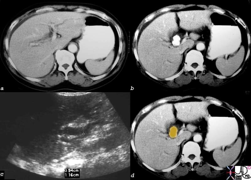
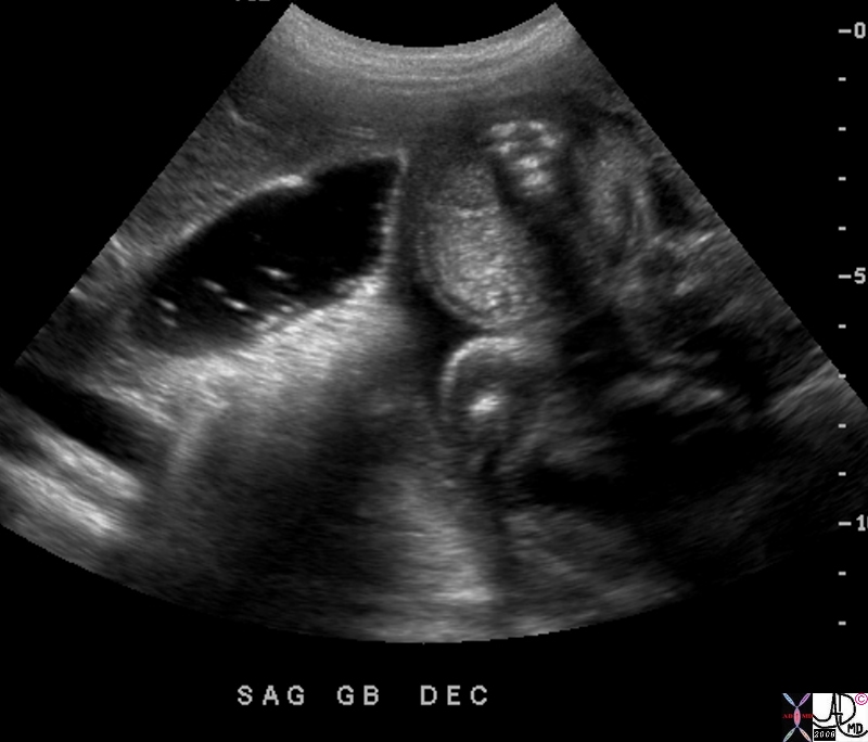
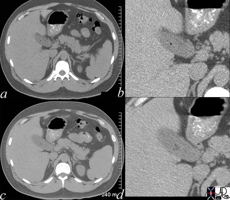
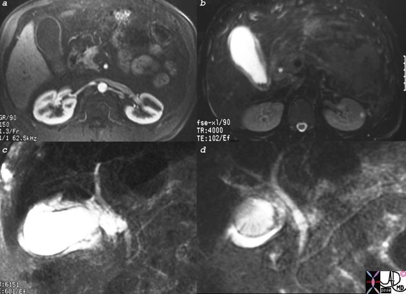
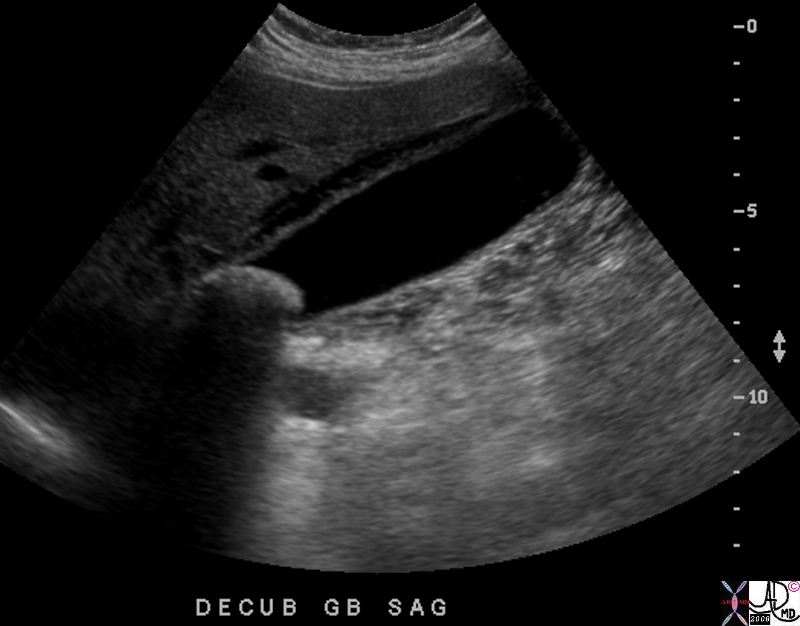
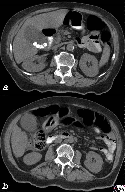
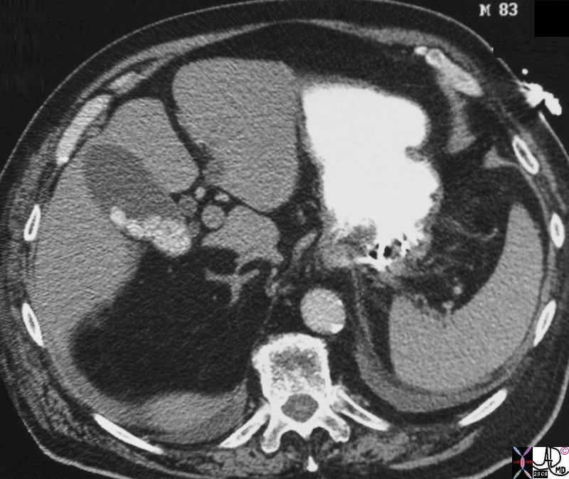
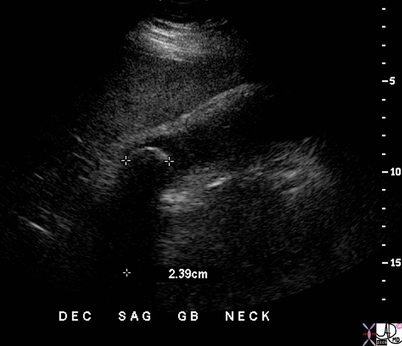
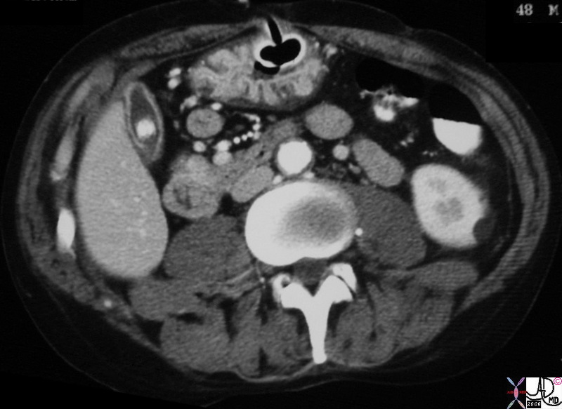
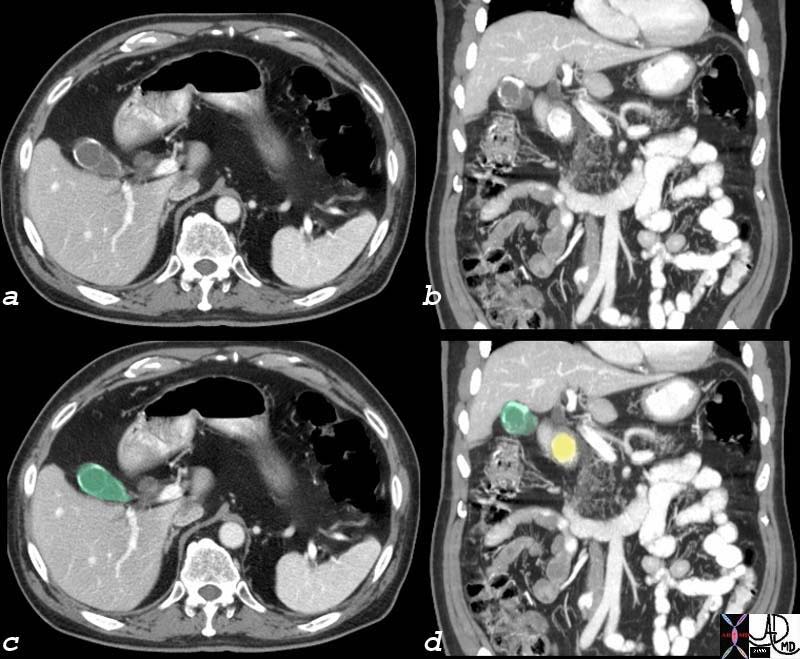
Mirizzi’s Syndrome – Large Stone in the Infundibulum Bile Duct Obstruction |
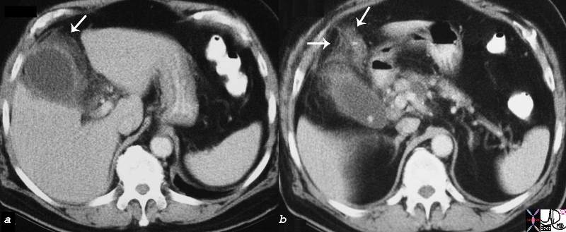
| 57year old male presents with RUQ pain fever, positive Murphy’s sign, elevated alkaline phosphatase. The CTscan in a shows mild intrahepatic bile duct dilatatio, while image b shows a 2.8cms stone impacted in the region of the infundibulum of the gallbladder. Image c is an ultrasound which shows dilated ommon hepatic duct (up to 1.2cms) while d shows an overlay in orange of the gallstone. At surgery the stone was noted to be impacted in an Hartmann’s pouch. The combination of acute cholecystitis with bile duct obstruction caused by mass effect of a stone in the base of the gallbladdercausing extrinsic compression is called Mirizzi’s syndrome.
gallbladder stone cholelithiasis cholecystitis bile duct obstruction Mirizzi’s syndrome USscan ultrasound CTscan CourtesyAshley Davidoff MD copyright 2008 82342c.8s |
Shape
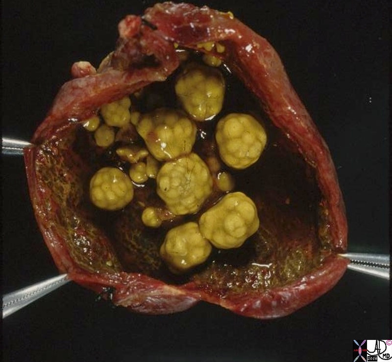 
Mulberry Shaped and Pebble Shaped |
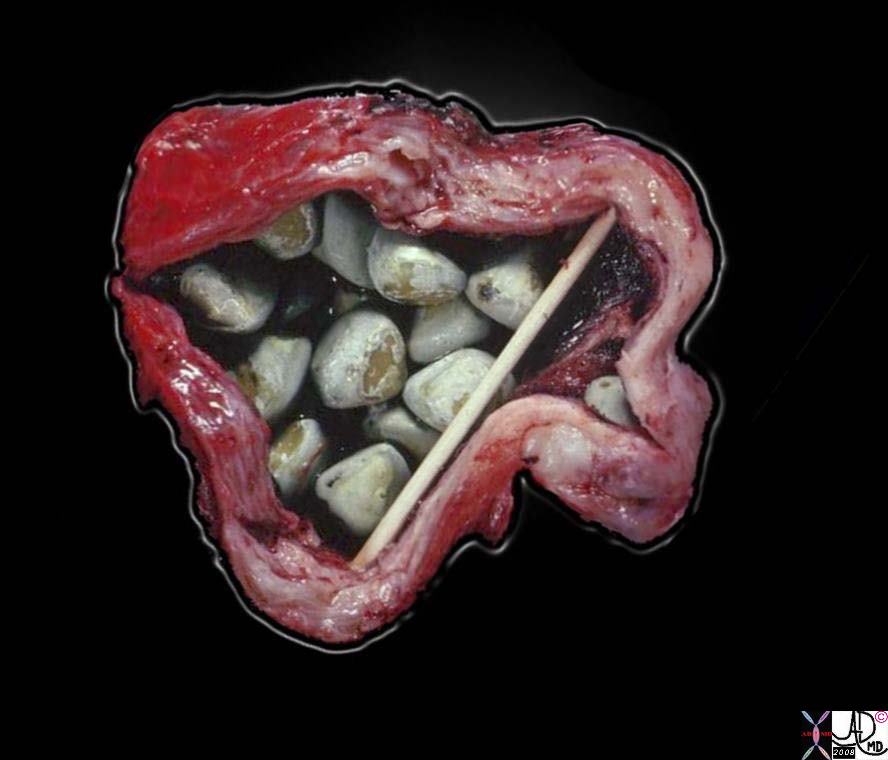
|
On the left is a photograph of a resected gallbladder opened longitudinally. The gallbladder is being held open by hemostats, as the inflammatory process made the wall thicker and stiffer than normal. Note that the cut face of the wall between the hemostats is about 3 – 4 mm thick. The dark red color is imparted by hemorrhage in the wall. The multiple gallstones are probably causing some degree of obstruction. On the right is a photograph of a resected gallbladder opened longitudinally. The gallbladder is being held open by sticks, as the inflammatory process made the wall thicker and stiffer than normal. Note that on the cut face the wall is about 1 cm thick. The color is pale tan-white due to fibrosis in the wall. The multiple gallstones are expected; it would be unusual not to see them.
11927.8s gallbladder cholecytitis grosspathology Courtesy Barbara Banner MD 11930.81s gallbladder chronic cholecystitis grosspathology Courtesy Barbara Banner MD
|
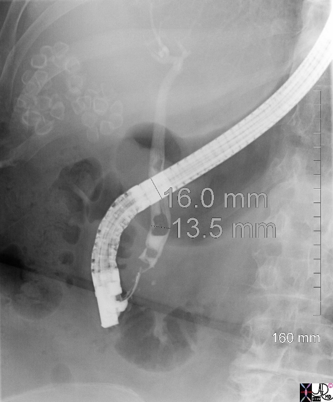
Faceted and Calcified Stones |
|
This ERCP shows gallstones are partially calcified around their rim and can be seen on their own, as happens about 10% of the time. Thety are faceted. 0026.8s gallbladder stones cholelithiasis multifaceted small bile duct choledocholithiasis ERCP Courtesy Ashley Davidoff MD copyright 2008 |
Position
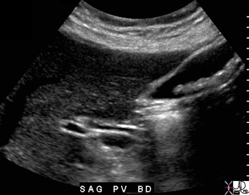
Dependant Position and Mobile
Small Pigmented Stones Lying Dependantly in the Gallbladder |
| 11921.8b05b018.8s gallbladder cystic duct gallstones cholelithiasis Davidoff Art copyright 2008 |

Mobile Stone |
| 46462c01s gallbladder moderaste size shadowing mobile stones cholelithiasis USscan ultrasound Courtesy Ashley Davidoff MD copyright 2008 |
Applied Biology
Stuck in the Neck
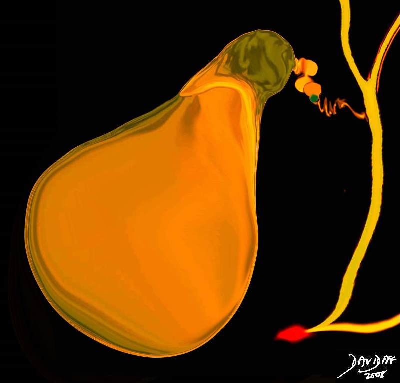
Stone Impacted in the Cystic Duct Causing Obstruction |
| 11921.8b05b019.8s gallbladder cystic duct gallstones cholelithiasis stone impacted in the cystic duct distended enlarged Davidoff Art copyright 2008 |
|
|
| 78305.8s gallstone large stone cholelithiasis impacted in the infundibulum does not move with decubitus fluid in the gallbladder fossa acute cholecystitis USscan ultrasound Courtesy Ashley Davidoff Md copyright 2008 |
Stuck in the Cystic Duct
Stuck in the Cystic Duct |
| 04766b05b04.35k.8s gallbladder cystic duct stone impacted obstruction enlarged gallbladder acute calculous cholecystitis Davidoff art copyright 2008 |
Character
Calcification
Dense “Hard” Calcification |
| 77728c01.8s gallbladder edematous wall distended gall bladder edema in the wall fluid in the fat surrounding the gallbladder indurated fat acute cholecystitis calcified stones cholelithiasis stones visible on CT scan CTscan Courtesy Ashley Davidoff MD copyright 2008 |
Dense Calcification Causing Echogenic Rim and Significant Shadowing |
| 00136 gallbladder stone large shadowing cholelithiasis imaging radiology USscan |
“Soft” Calcification |
| 26073.8s gallbladder stones moderate calcification soft calcification gallstone pancreatitis dx cholelithiasis Courtesy Ashley Davidoff MD copyright 2008 |
Soft Calcification – No Strong Rim and Texture of Matricx is Visualized8mms all Aligned |
| The echogenic nature of these stones with no strong echogenic front wall is more reminiscent of a cholesterol stones or mixed stones.
76040b01 gallbladder stones cholelithiasis bile duct normal portal vein liver vein USscan Courtesy Ashley DAvidoff MD |
Rim Calcification Small Stones have Homogeneous |
| 30836.8s gallbladder calcified cacalcification varied size stones cholelithiasis complex stones chronic cholecystitis CTscan Courtesy Ashley DAvidoff Md copyright 2008 |
|
Large Laminated Gallstone |
| 82151.8s 51f obesea gallbladder stone large calcified lamellated cholelithiasis CTscan Courtesy Ashley DAvidoff MD copyright 2008 |
Fat
Obesity and Cholesterol Stone |
| 82205.8s 82206.8s 54 female mildly enlarged non tender gallbladder no abnormality seen on CTscan but large stone seen on USscan consistent with cholesterol stone isodense on CT shadowing on ultrasound atony obese Courtesy Ashley Davidoff MD Copyright 2008 |
Cholesterol Stone |
| 25055 Courtesy Ashley Davidoff MD code pancreas fx cyst gallbladder + fx filling defect + fx floating + dx cholesterol cholelithiasis + imaging radiology CTscan C+ |
Mixed
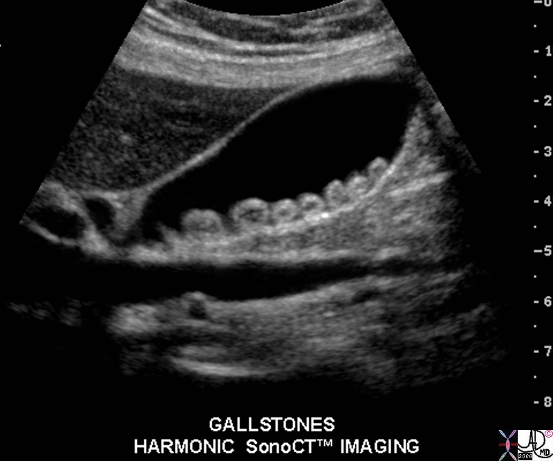
Echogenic but Reduced Calcification without Strong Shadows |
| 1994H5~1.8s gallbladder stones cholelithiasis hypoechoic centre harmonic sonoCT normal wall Courtesy Philips Medical Systems copyright 2008 |
Air
Floating and Gas Filled |
| 26540 gallbladder + fx filling defects + fx air + fx floating + dx cholelithiasis + imaging radiology CTscan C- |
|
Subtle Character Changes |
| 70269c02 gallbladder air methane nitrogen gas cholesterol stone isodense with bile fat subtle change CTscan |
Applied Biology
The Trouble Stones Cause
Complication of Gall Stone Disease
Acute Cholecystitis
Acute Cholecystitis |
| 00401.1cs 67Male right upper quadrant pain RUQ pain positive Murphy’s sign gallbladder gallbladder fossa thickened linear lacy thickening gall stones calculi calculous large stone in the infundibulum shadowing multiple small stones cholelithiasis cholecystitis acute cholecystitis acute calculous cholecystitis USscan ultrasound Courtesy Ashley Davidoff MD copyright 2008 |
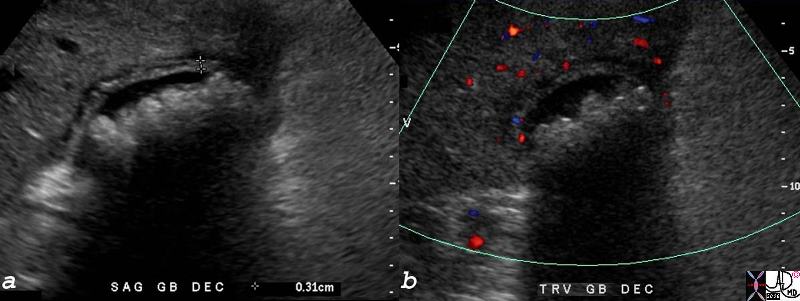
Gallbladder Filled with Stones |
| 77744c01.8s gallbladder distended enlarged pericholecystic fluid fluid in the gallbladder fossa wall thickened hyperemic almost completely filled with stones shadowing cholellithiasis hypervascular gbf acute cholecystitis USscan ultrasound color flow doppler Courtesy Ashley Davidoff MD copyright 2008 |
Acute Cholecystitis with Transmural Induration |
|
16203c01.8s gallbladder edematous wall hyperemic wall distended gall bladder edema in the wall fluid in the fat surrounding the gallbladder indurated fat acute cholecystitis stones visible on CT scan CTscan Courtesy Ashley Davidoff MD copyright 2008 |
Complications
Chronic Cholecystitis
Chronic Cholecystitis |
|
This is a photograph of a resected gallbladder opened longitudinally. The gallbladder is being held open by sticks, as the inflammatory process made the wall thicker and stiffer than normal. Note that on the cut face the wall is about 1 cm thick. The color is pale tan-white due to fibrosis in the wall. The multiple gallstones are expected; it would be unusual not to see them. 11930.81s gallbladder chronic cholecystitis grosspathology Courtesy Barbara Banner MD |
Chronic Cholecystitis, Hyperemic Wall, Small Contracted Gallbladder |
| 30701.8s stone cholelithiasis hyperemic wall small contracted chronic cholecytitis stomach gastrostomy redundant mucosa CTscan Courtesy Ashley Davidoff MD copyright 2008 chronic cholecytitis |
Chronic Cholecystitis Enhancing Wall |
| 16239c.8s gallbladder stone cholelithiasis wall thickened thick wall enhancing wall dx chronic cholecystitis bile duct cholangiocarcinoma MRI with and without contrast |
|
Chronic Cholecystitis – Hour Glass Focal Fibrosis |
| 11530.81s gallbladder shape deformity filling defect figure of eight defect cholelithiasis chronic cholecystitis oral cholecystogram historical Courtesy Ashley DAvidoff MD copyright 2008 |
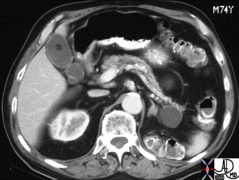
Gallstone Pancreatitis
Gallstone Pancreatitis |
| 05265 Courtesy Ashley Davidoff MD code pancreas fx air fx fluid cystic fx mass dx necrotizing pancreatitis code gallbladder fx stones dx cholelithiasis code lung pleura fx effusion dx sympathetic effusion dx gallstone pancreatitis complicated by gangrene necrosis possible infection imaging radiology CTscan code acute inflammation |
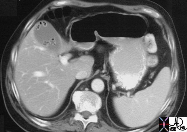
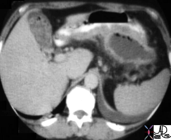
Porcelain Gallbladder
Porcelain Gallbladder with Calcified Stone |
|
In this CTscan calcification of the wall of the gallbladder (green) is associated with a large calcified stone (yellow) that appears to be impacted in the neck. Cholesterol stones are usually isodense with the bile but in this case it has calcified. The large size of the stone suggests its cholesterol nature in this case. 76225c01 gallbladder wall calcification calcified porcelain gallbladder premalignant CTscan Courtesy Ashley Davidoff MD |
Gallstone Ileus
Gallstone Ileus |
| 31127c01.8s gallbladder stones air adherent to duodenum small bowel dilatation calcification stone gallstone ileus CTscan Courtesy Ashley DAvidoff copyright 2008 |
| Gallstone Ileus |
| 31127c02.8s gallbladder stones air adherent to duodenum small bowel dilatation calcification stone gallstone ileus CTscan Courtesy Ashley DAvidoff copyright 2008 |
Gallbladder Carcinoma
| Local Invasion and Separate Liver Mestastasis |
| 17280c02b01.8s gallbladder liver mass local invasion cholelithiasis metastasis carcinoma primary gallbladder gallbladder fossa Courtesy Ashley Davidoff MD copyright 2008 |
| Local Invasion Bile Duct Obstruction Peritoneal Involvement |
| 17280c03.8s gallbladder liver mass local invasion cholelithiasis metastasis carcinoma primary gallbladder gallbladder fossa invasion into gastrohepatic ligament bile ducts obstructed ascites Courtesy Ashley Davidoff MD copyright 2008 |
References
Aspinall By Richard J. Aspinall, Simon D. Taylor-Robinson, Joanna Cameron Mosby’s Color Atlas and Text of Gastroenterology and Liver Disease Published by Elsevier Health Sciences, 2001
Rosenthal Principles and practice of geriatric surgery Ronnie A. Rosenthal, Michael E. Zenilman, Mark R. Katlic Published by Springer, 2001
Faye Maryann Lee, MD e Medicine Cholellithiasis Last updated June 2006