
The Common Vein Copyright 2008
Alok Anand
Definition
Chronic cholecystitis is a repetitive inflammation of the gallbladder. Unlike acute cholecystitis, it is unclear whether gallstones are involved in the process and its subsequent clinical presentation of pain, although stones are present in nearly 90% of cases.
The patient may have an indolent RUQ pain, or biliary colic. It is similar to thate of acute cholecystitis, and is accompanied by nausea, vomiting and frequently intolerance of fatty foods.
There are important structural changes associated with chronic cholecystitis. Proliferation of immune cells, fibrosis, and proliferation of the mucosal epithelium. Mucosal epithelium often gives rise to the formation of deep crypts within and through the wall, often called Rokitansky-Aschoff sinuses.
Whereas acute cholecystitis often presents with fever, in chronic cholecystitis, patients are usually afebrile and do not usually have an elevated white cell count. The may have localized tenderness over the gallbladder (Murphy’s Sign). The inflammatory processes may lead to modestly abnormal liver function tests, namely high transaminase levels.
Increased dystrophic calcification may yield an accumulation of mineral called a porcelain gallbladder, which is associated with carcinoma.
In addition, obstruction and derangements of the cellular function from chronic inflammation may result in an accumulation of clear secretions, referred to as hydrops of the gallbladder.
It may result in a variety of complications similar to that of acute cholecystitis, namely bacterial superinfection, perforation with abscess formation, or rupture with bile peritonitis.
Diagnosis is often with ultrasound, and can be confirmed with CT imaginge. Findings include stones in the gallbladder. There is often a thickened gallbladder wall. In advanced cases, the wall may even be contracted.
 Chronic Cholecystitis Chronic Cholecystitis |
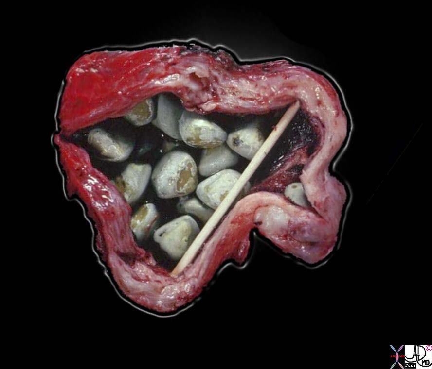
| This is a photograph of a resected gallbladder opened longitudinally. The gallbladder is being held open by sticks, as the inflammatory process made the wall thicker and stiffer than normal. Note that on the cut face the wall is about 1 cm thick. The color is pale tan-white due to fibrosis in the wall. The multiple gallstones are expected; it would be unusual not to see them.
11930.81s gallbladder chronic cholecystitis grosspathology Courtesy Barbara Banner MD |
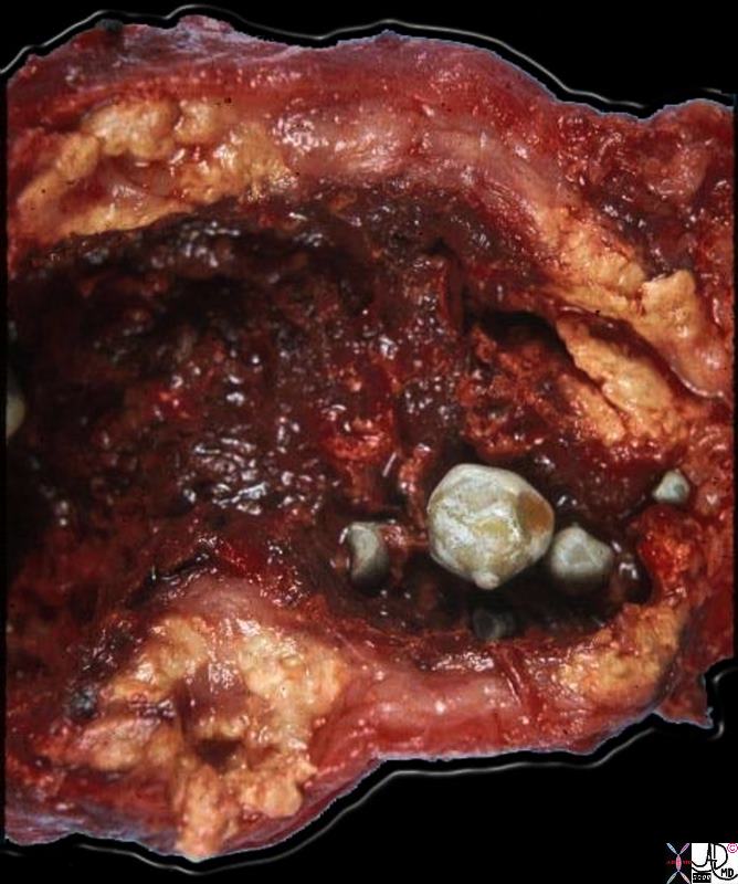
Hemorhagic Cholecystitis – Acute on Chronic |
|
This gallbladder was opended longitudinally to show the lumenal surface. Four gallstones are present. The mucosal surface looks hemorrhagic, indicative of active inflammation. The most striking aspect of this gallbladder is the marked thickening and yellow color of its walls. This is due to extrusion of bile into the wall during inflammation when the integrity of the mucosa is interrupted. Once in the tissues of the wall, the bile triggers an intense inflammatory reaction, and a response by histiocytes, which phagocytose the bile and attempt to break it down. The marked accumulation of lipids and their break-down products imparts the yellow color. The significance of this variant of chronic cholecystitis is that it mimicks carcinoma by the way it thickens the wall. The radiologist, the surgeon, and even the pathologist may think this is carcinoma until the microscopic sections are examined. 11937b01.8s gallbladder dx cholelithiasis grosspathology Courtesy Barbara Banner MD |
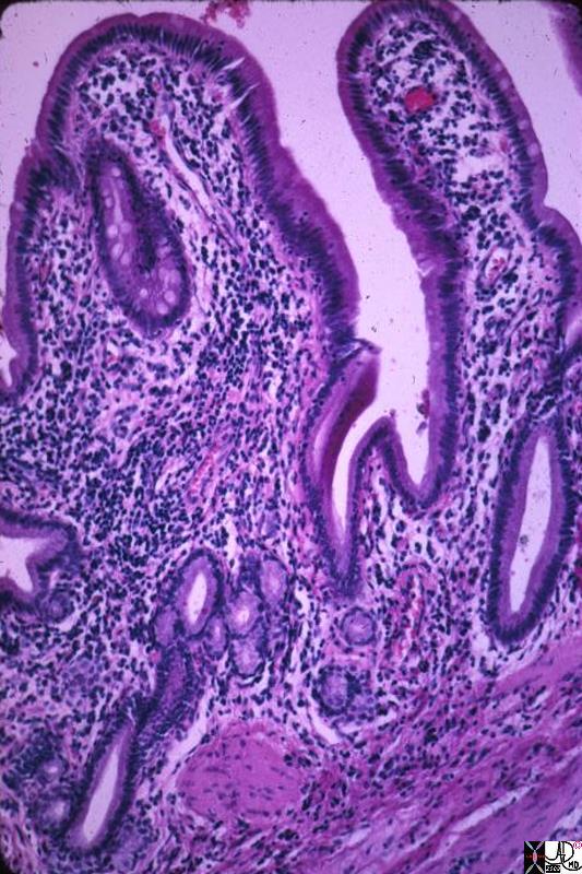
Metaplasia – Sign of Chronic Inflammation |
| This medium power photomicrograph shows gallbladder mucosa with an inflammatory infiltrate of mononuclear cells. The lumen is the clear area, and a tiny part of muscular layer can be seen at the opposite side of the picture. Look carefully and you will find one area near the lumen with goblet cells, like intestinal epithelium. This is intestinal metaplasia. Deeper in the mucosa near the muscular layer is a cluster of a few glands which do not have a tall columnar epithelium. They resemble gastric glands, and in fact this is gastric metaplasia. Metaplastic epithelium occurs frequently throughout the biliary and gastrointestinal tracts in chronic inflammatory conditions. In the gallbladder it has no significance other than being part of the changes due to chronic inflammation.
11932.8s gallbladder chronic cholecystitis histopathology Courtesy Barbara Banner MD |
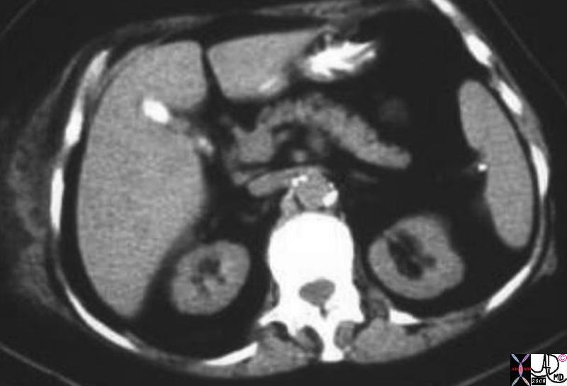
Small Gallbladder Filled with Stones |
| 02118.8s gallbladder small stone filled contracted chronic cholecytitis CTscan Courtesy Ashley Davidoff MD copyright 2008 |
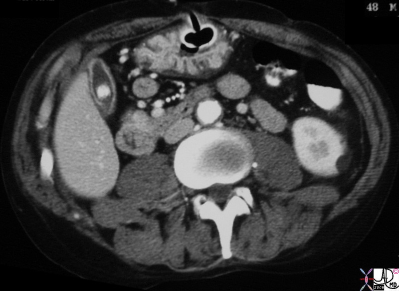
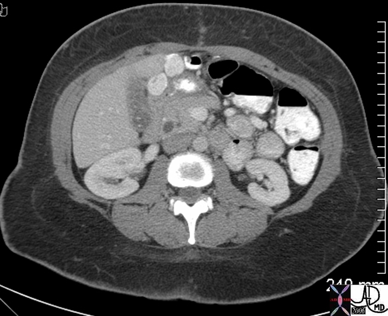
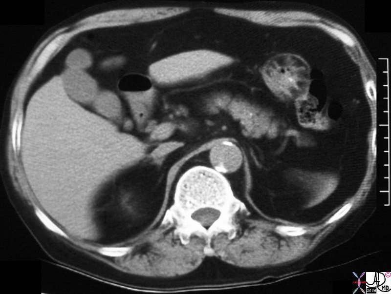
Chronic Cholecystitis, Hyperemic Wall, Small Contracted Gallbladder |
| 30701.8s stone cholelithiasis hyperemic wall small contracted chronic cholecytitis stomach gastrostomy redundant mucosa CTscan Courtesy Ashley Davidoff MD copyright 2008 chronic cholecytitis |
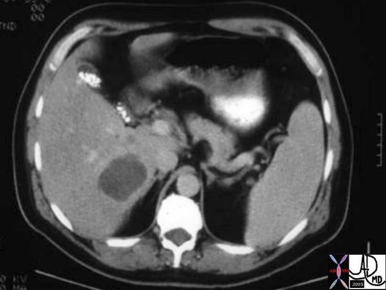
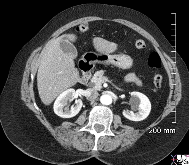
Small Gallbladder Filled with Stones with Liver Abscess |
| 02249 liver fx mass infection abscess dx diverticulitis gallbladder gallstones dx cholelithiasis CTscan Courtesy Ashley Davidoff MD |
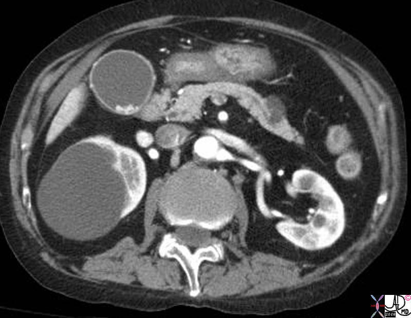
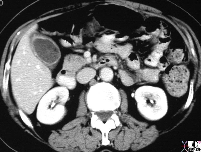
Chronic Cholecystitis, Hyperemic Wall, Prominent Arterioles, Distended Lumen |
| 39408.8s gallbladder calcified stones cholelithiasis hyperemic wall cystic artery branches hyperemic mucosa and submucosa ‘kidney renal cyst pancreas chronic cholecytitis CTscan Courtesy Ashley Davidoff MD copyright 2008 |
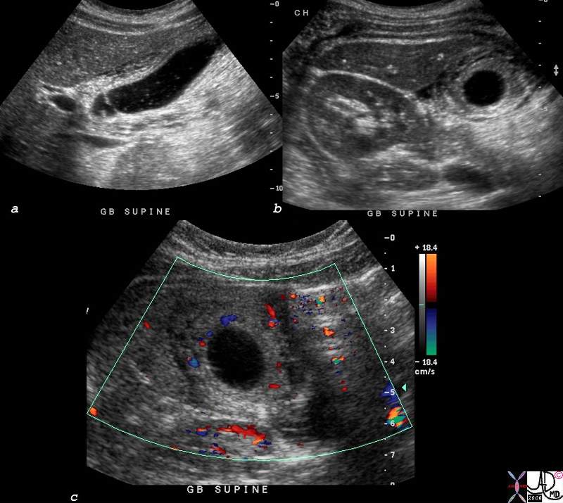
Acute on Chronic Cholecystitis |
| 81983c01.8s 31 F pw abdominal pain chronic cholecytitis cholelithiasis at path fluid was gelatinous green cholesterol choleliths measuring up to 6mm focal congestion of mucosa cystic duct occluded with 9mm mulberry cholesterol stone gallbladder thick wall hyperemic cholesterol crystals complex fluid in the gallbaldder fossa GBF dx acute on chronic cholecystitis USscan ultrasound color flow doppler Courtesy Ashley Davidoff MD copyright 2008 |
Tiny Calcified Floaters |
| 45092 45092b02 gallbladder fx cholelithiasiscompletely occupying gallbladder lumen CTscan Courtesy Ashley Davidoff MD |
Tiny Calcified Cholesterol Floaters |
| 25306.8s small cholesterol crystals stones floaters hyperemic wall chronic cholecytitis gas air cholelithiasis CTscan Courtesy Ashley Davidoff MD copyright 2008 |
Layering Hyperdense |
| 78352.8s obese female gallbladder layering hyperdense stones cholelithiasis hyperemic wall hyperemic mucosa and submucosa chronic cholecystitis CTscan Courtesy Ashley Davidoff MD copyright 2008 |
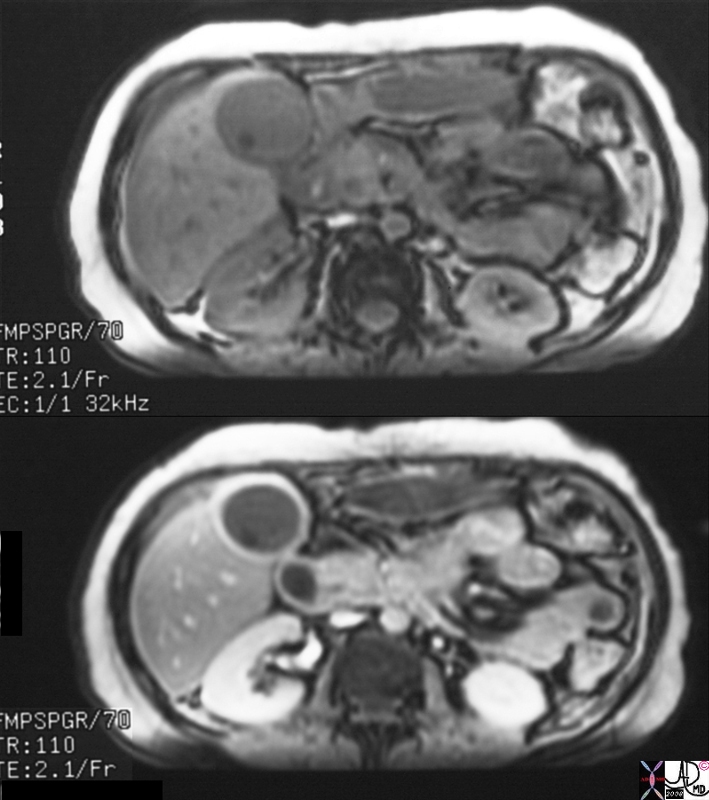
Chronic Cholecystitis Enhancing Wall |
| 16239c.8s gallbladder stone cholelithiasis wall thickened thick wall enhancing wall dx chronic cholecystitis bile duct cholangiocarcinoma MRI with and without contrast |
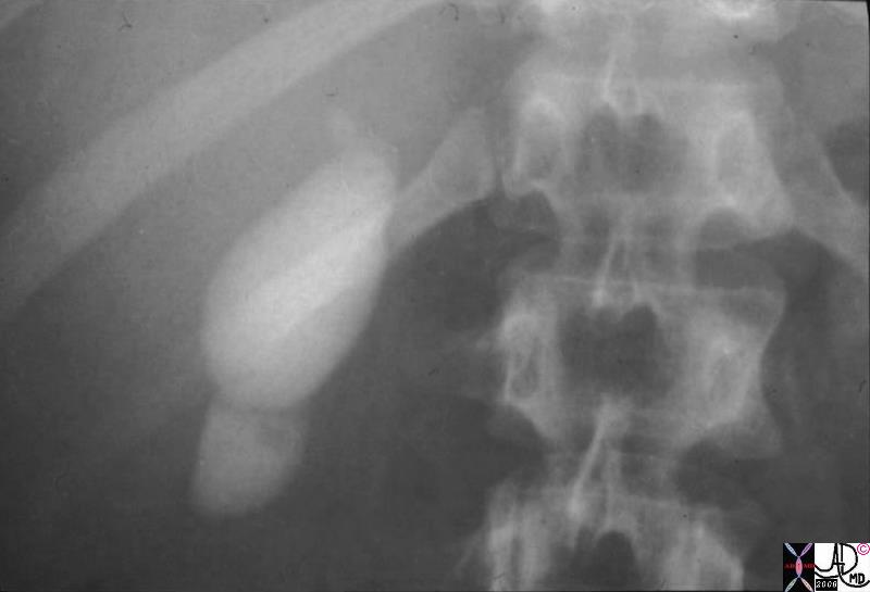
Figure of 8 Chronic Cholecystitis |
| 04731.8s gallbladder fibrosis across the fundus filling defect chronic cholecystitis OCG historical stricture Courtesy Ashley Davidoff MD copyright 2008 |
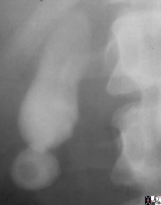
Figure of 8 Chronic Cholecystitis |
| 11530.81s gallbladder fibrosis across the fundus filling defect chronic cholecystitis cholelithiasis stone in fundus OCG historical stricture Courtesy Ashley Davidoff MD copyright 2008 |
Chronic Cholecystitis |
| 31285.8s 64 male gallbladder fibrosis across the fundus filling defect chronic cholecystitis cholelithiasis stone in fundus Ctscan figure of eight stricture Courtesy Ashley Davidoff MD copyright 2008 |
|
Chronic Cholecystitis, Hyperemic Wall, Small Contracted Gallbladder |
| 30701.8s stone cholelithiasis hyperemic wall small contracted chronic cholecytitis stomach gastrostomy redundant mucosa CTscan Courtesy Ashley Davidoff MD copyright 2008 chronic cholecytitis |
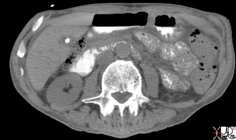
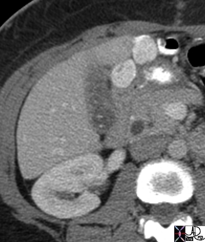
Small Contracted Gallbladder with Stone and Thick Wall |
| 81916.8s gallbladder stone cholelithiasis thick wall thickened wall small contracted chronic cholecystitis CTscan Courtesy Ashley Davidoff MD copyright 2008 |
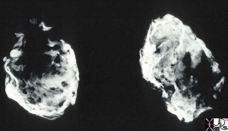
Porcelain Gallbladder – Variant of Chronic Inflammation |
| This is a specimen xray of a resected gallbladder. The walls were thin, fibrotic and heavily calcified, accounting for the radiodense (white) areas on the radiograph. This variant of chronic cholecystitis is a risk factor for development of carcinoma.
11939.8s gallbladder porcelain gallbladder X-ray Courtesy Barbara Banner MD |
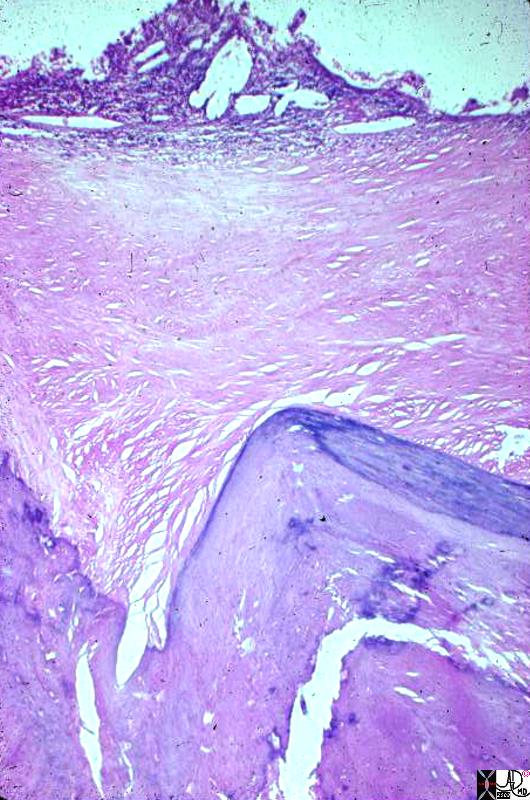
Porcelain Gallbladder – Variant of Chronic Inflammation |
| This photomicrograph at low power shows most of the full thickness of the wall of a gallbladder. The lumen is the clear area at one side of the picture. The pale homogeneous area down the middle is fibrous replacement of the wall. The little nubbin of tissue and cholesterol clefts along the lumen represents what is left of the mucosa. The darker, zig-zag structure at the side opposite the lumen represents where the gallbladder wall is calcified. This calcification and fibrosis of the entire gallbladder wall is the diagnostic feature of porcelain gallbladder.
11940.8s gallbladder porcelain gallbladder histopathology Courtesy Barbara Banner MD |