
Gallbladder
The Common Vein Copyright 2008
Introduction
The normal gallbladder in the fasting state contains about 50-70mls of bile and measures 8-10 cms in length, by 3-4cms in diameter using ultrasound. Because of its ellipsoid shape the approximation for gallbladder volumecan be estimated by using the formula of length X width X depth X .5cms.
Common Causes
Enlargement is usually caused by obstruction by calculi, which typically become lodged in the neck of the gallbladder in the cystic duct or more distal obstructions, in the common bile duct. Neoplasm of the bile duct, metastases to porta hepatis nodes and pancreatic carcinoma, are not uncommon causes of a distended gallbladder. Clot in the biliary tree may also cause obstruction.
Less commonly, the gallbladder can become distended due to atony. This is commonly seen in diabetic neuropathy but is also caused by aging and cholestasis. Prolonged cholestasis is commonly seen in severely ill ICU patients who have gallbladders that are unstimulated by CCK. Decreased contractility can also result from impaired vagal nerve function, due to iatrogenic or traumatic severing of the vagal innervations to the gallbladder.
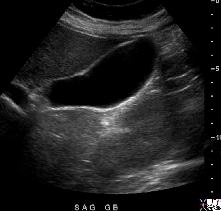
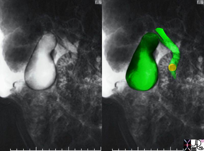
Normal Size of the Fasting Gallbladder on Ultrasound |
| The gallbladder in the fasting state measures 8-10 cms in length and 3-4cms in diameter. In this instance it measures 10 by 3.5cms.
82021.8s gallbladder normal anatomy size shape position character width 3.5cms length = 10cms pear shape pyriform shape fundus body neck fundus anterior neck posterior USscan ultrasound Courtesy Ashley Davidoff MD copyright 2008 pear shape |
The wall is normally 2-3mm thick in the fasting state. It is only about 1mm thick in the neonate.
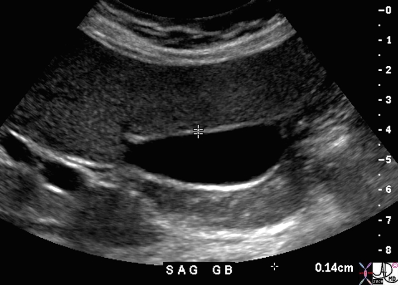
Wall Thickness by Ultrasound |
| 82427.8s gallbladder wall normal size thickness USscan ultrasound Courtesy Ashley DAvidoff MD Copyright 2008 |
Applied Biology
The size of the gallbladder is one of the more important determinants in the evaluation of gallbladder disease, but as you will learn there is not always a black and white answer as to whether the gallbladder is enlarged or not.
The size of the gallbladder cannot be assessed by clinical examination since the gallbladder lies under the liver and only a small portion of the fundus is exposed. Therefore under normal circumstances it cannot be palpated and is only palpable when it is significantly enlarged. For this reason, assessment of the size of the gallbladder requires the use of an imaging technique. This is best accomplished by ultrasound, since the true long axis and short axis can be defined by the ultrasonographer who is able to angle the transducer along the appropriate axis. The gallbladder is normally positioned at an angle that is off the horizontal, sagittal and coronal planes. The fundus is located relatively inferiorly, while the neck is positioned more medially and superiorly. Thus by the nature of the transverse imaging of CT and usual orthogonal imaging of MRI, the true axis is not routinely defined by these methods. Although nuclear imaging can also assess the size of the gallbladder accurately, other factors such as wall thickness and probe tenderness cannot be evaluated. Thus when assessment of the size of the gallbladder, and associated features of wall thickness and probe tenderness is required, ultrasound becomes the procedure of choice.
From a practical and clinical standpoint, the clinician who suspects an enlarged gallbladder is really asking whether there is increased pressure in the gallbladder. The imaging technology uses size as an indirect measure of the intraluminal pressure since we would otherwise have to use invasive techniques to assesss the pressure directly. Since there are situations where the gallbladder loses its tone (atonic or hypotonic gallbladder as a result of aging process or diabetes) it can enlarge without the ever important increase in pressure. On the other hand there are situations when the gallbladder wall is fibrotic (chronic cholecystitis) and this results in a small contracted gallbladder which cannot dilate when the luminal pressure rises.
Multiple factors therefore have to be taken into consideration when assessing the size of the gallbladder.
The key to assessing gallbladder size is a transverse view through the body. This requires a transverse image that is orthogonal to the long axis of the gallbladder. In this view as shown below the transverse (right side to left side) dimension is larger than the anteroposterior (A-P) dimension. This is normal. It implies that that the luminal pressure is less than the pressure exerted by the liver and so the liver flattens the anterior wall. Sometimes the normal gallbladder has a larger A-P dimension depending on the external forces exerted on it. It is not usually round. The normal largest dimension in the transverse should not be greater than 4-5cms.
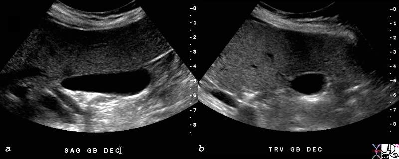
The Shape of the Gallbladder in Transverse Section |
| 82428c01.8s gallbladder small normal transverse oval shape normal anatomy USscan ultrasound copyright 2008 Courtesy Ashley DAvidoff MD |
Sometimes the structures on the free wall of the gallbladder compress it. We can thus infer that gallbladder pressure is lower than the structures around it and is therefore normal.
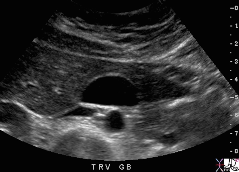
Shape Reflecting Size |
|
In this very thin patient the posterioor wall is flattened by posterior structures. The presure in the gallbladder therefore has to be low. 82418.8s 38F normal gallbladder pressure size normal anatomy Courtesy Ashley Davidoff MD copyright 2008 |
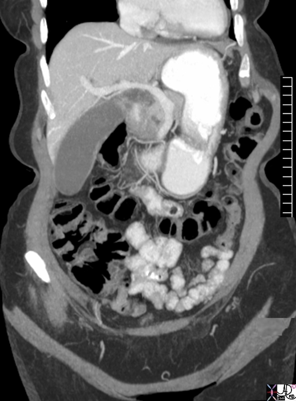
Elongated but not Dilated – The Aging Gallbladder |
| The CT is also from an elderly asymptomatic female (from the perspective of the gallbladder) whose gallbladder measures 3.5cms in the short axis and 11cms in the long axis. The latter measurement is considered abnormal while the former is normal. This zuchini shaped gallbladder is probably normal and reflective of the patients age. An ultrasound would be far more specific, since wall thickness, probe tenderness, and true long axis and short axis measurements could be made.
76764.8s elderly femal gallbladder elongated sausage shape zuchini shape transverse dimension 4.5 cms longitudinal dimension 12.5cms aging gallbladder vs enlarged? CTscan Courtesy Ashley DAvidoff MD copyright 2008 |
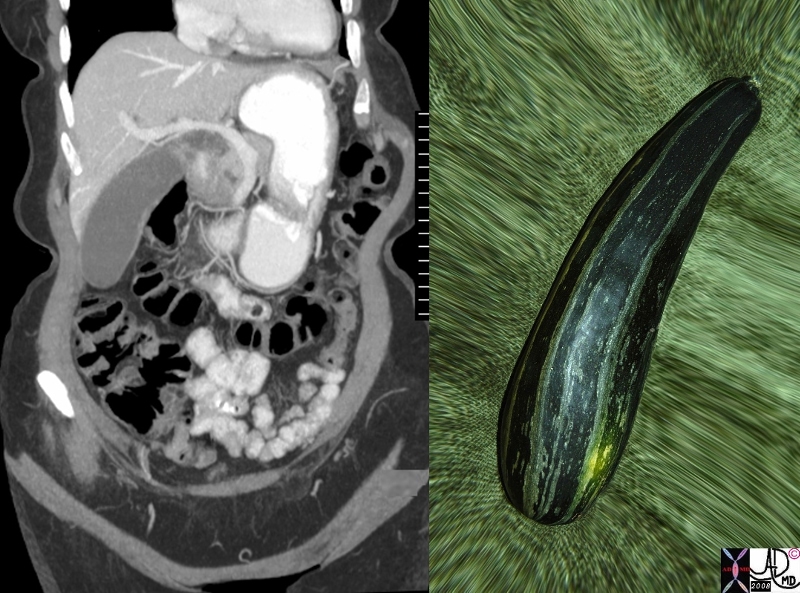
Zuchini (Italian) Gallbladder |
|
The elongated shape of the gallbladder without being wide is remoiniscent of the Italian Zuchini in thi as76764c.81s elderly female gallbladder elongated sausage shape zuchini shape transverse dimension 4.5 cms longitudinal dimension 12.5cms aging gallbladder vs enlarged? food in the body CTscan Courtesy Ashley DAvidoff MD Davidoff art Davidoff photography copyright 2008 |
The Large Gallbladder
The large galllbladder is defined as a gallbladder that exceeds 10cms in the long axis and 4cms in the short axis. Although it is not definitive, the transverse measurement of the gallbladder is more specific in the evaluation of size. An elongated gallbladder can be normal, particulalrly in the elderly. The shape of the gallbladder becomes more rotund when it is enlarged. The gallbladder is normally ovoid in shape in the transverse plane. With enlargement it becomes round which is a fairly reliable criterion for a dilated gallbladder.
Gallbladder enlargement is not necessarily abnormal. In aymptomatic patients, enlargement can occur as a normal variant in people with asthenic builds, in older patients and in diabetics (atonic). It is suggestive of cholestasis in patients who are on a prolonged fast, and those on hyperalimentation. An atonic or hypotonic ileus of the gallbladder is seen in patients following abdominal surgery, patients with GI infections, hepatitis, pancreatitis, and bowel ileus, all of which can cause asymptomatic enlargement. In these patients there is no gallbladder tenderness, the wall is normal , but there may be thickened bile (sludge) in the lumen.
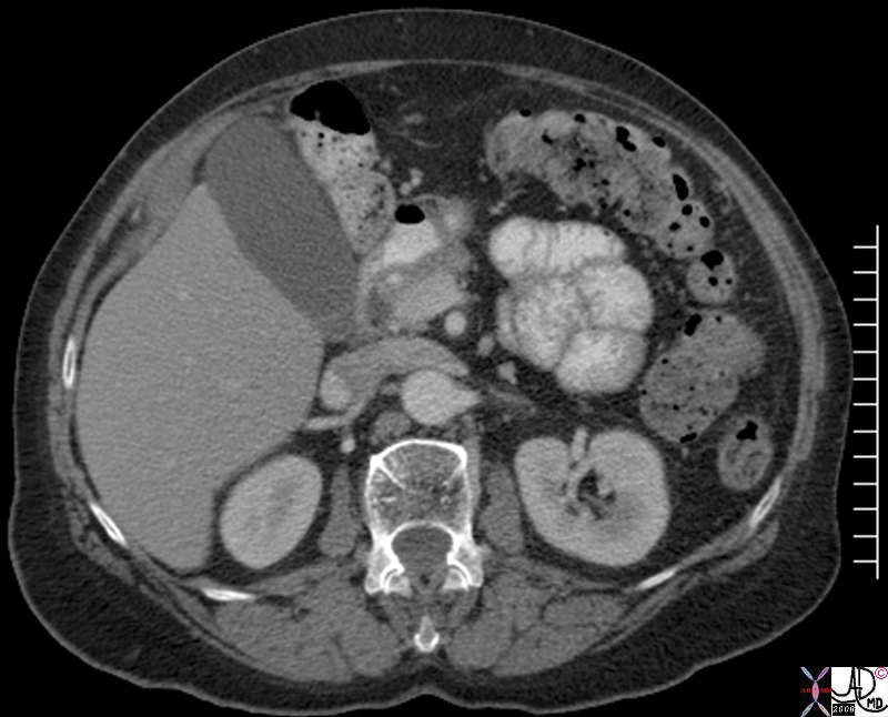
Gallbladder Upper limits Normal in Size |
|
The 78 year old female is asymptomatic from the perspective of the gallbladder. It measures by CT on this cut 9.5cms in long axis and 3.5cms in short axis. This measurement is upper limitsnormal and probaly reflects age related change. 82156.8 78 year old female patient No gallbladder symptoms gallbladder normal atonic aging CTscan Courtesy Ashley Davidoff copyright 2008 |
Gallbladder enlargement is most often caused by mechanical abnormalities and usually by a stone that is impacted in the infundibulum, cystic duct or common bile duct. (CBD). Cystic duct or infundibular obstruction by a stone is the most common cause. If this results in inflammation then acute cholecystitis results.
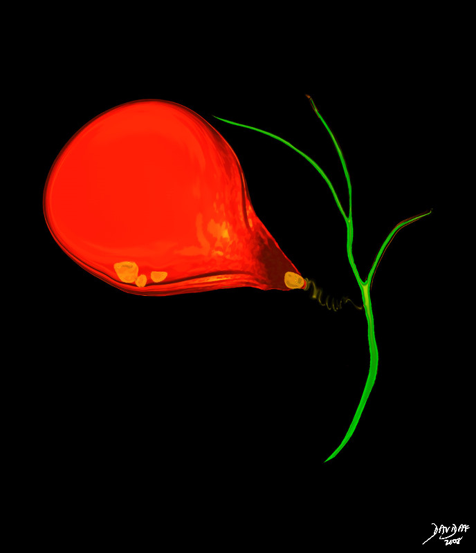
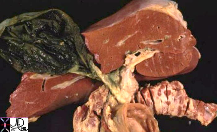
Acute Cholecystitis |
|
In this artistic rendition, a small stone has become impacted in the infundibulum of the gallbladder, resulting in obstruction and acute cholecystitis. 04766b08.8s gallbladder stones cholelithiasis acute cholecystitis distended enlarged bile ducts Davidoff art copyright 2008 |
A biliary stone may also becpome impacted in the common bile duct in which case dilatation of the bile ducts as well as the gallbladder results.
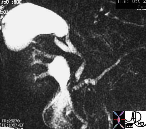
Choledocholithiasis |
|
The MRCP performed using T2 weighted FSE technique shows a CBD stone associated with dilatation of the CBD, cystic duct and the gallbladder. 17083c02.3k.8s 82 female presents with obstructive jaundice gallbladder duct enlarged distended distension dilated dilatation choledocholithiasis stone obstruction obstructive jaundice MRI T2 weighted Courtesy Ashley Davidoff MD copyright 2008 |

Acute Calculous Cholecystitis wit Gallbladder Wall Edema and Hyperemia |
| 72365c01 gallbladder wall fluid in gallbladder fossa positive sonographic Murphy’s sign inflammation mechanical obstruction infection hyperemia hypervascularity dx acute calculous cholecystiis Davidoff MD |
Obstruction by pancreatic cancer, or less commonly an ampullary tumor will result in an enlarged gallbladder. Other causes include sclerosing cholangitis when the common bile duct and less commonly the cystic duct are stenotic.
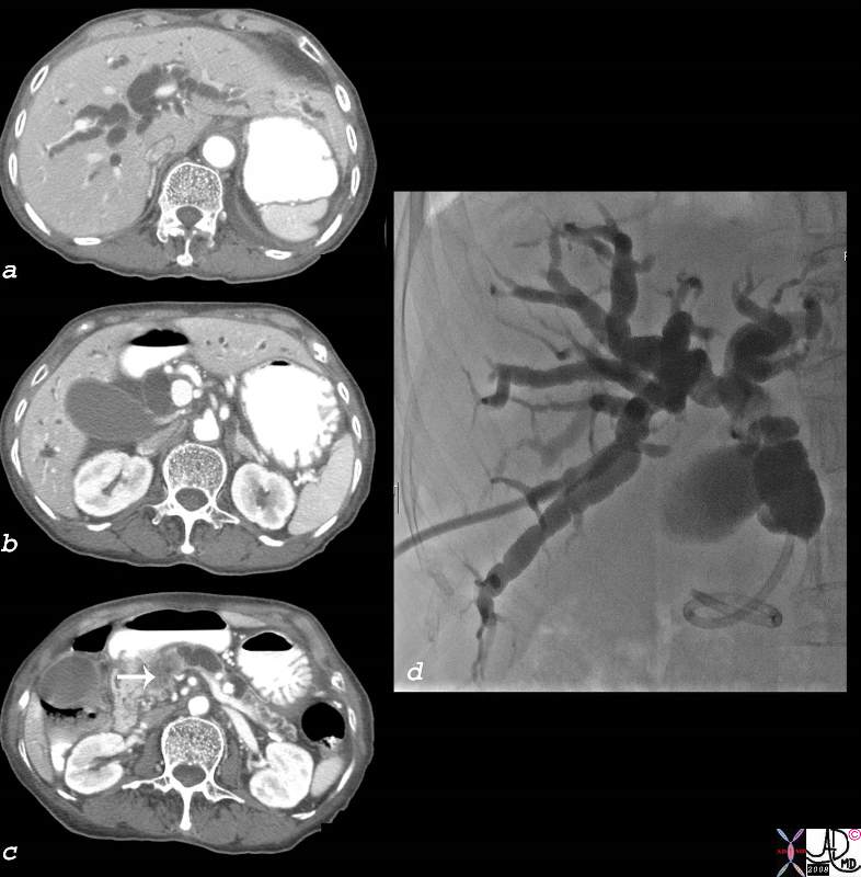
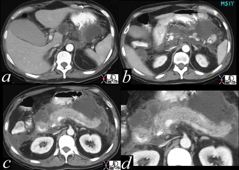
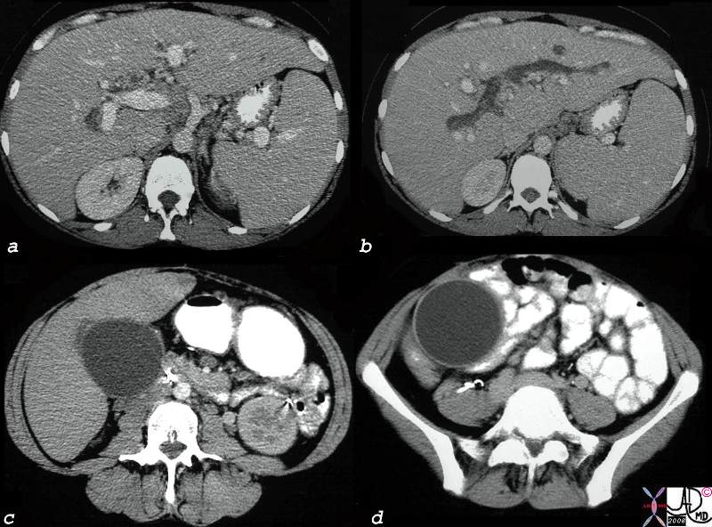
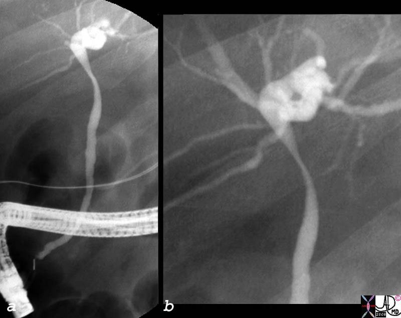
Courvoisier Gallbladder |
|
The patient presented with painless jaundice and a palpable gallbladder. (Courvoisier sign) The epicentre of the collage of images is in image c where the arrow points to a mass in the head of the pancreas that has caused obstruction of the pancreatic duct (seen upstream from the mass in c ), gallbladder distension (a) and bile duct dilatation (a,b, &d) A percutaneous biliary drainage catheter has traversed the obstruction and is in the duodenum that will allow for decompression. (d) 44005c01.8s elderly patient presents with painless jaundice Courvoisier gallbladder distended enlarged dilated pancreatic duct pancreas mass pancreatic carcinoma CTscan percutaneous cholangiogram PTC Courtsy Ashley DAvidoff MD copyright 2008 bile duct |
Acalculous cholecytitis is a relatively common cause of gallbladder dilatation. It is an advanced form of cholestasis where inflammatory changes supervene as a complication of prolonged and advancing cholestasis.
RUQ Pain, Distended Gallbladder, Indurated Fat – Acalculous Cholecystitis |
|
The CT iamges are from a an ICU patient who presented with RUQ pain and fever. The CTscan shows a distended gallbladder, no calcified stones, thickening and hyperemia of the wall, with stranding in the fat surrounding the gallbladder. Although this could still be calculous cholecystitis caused by an impacted stone the clinical setting is more likely to be of an acalcalous nature. Prior to placing a cholecystostomy tube US scan should be performed to exclude calculi. 77717c02s right upper quadrant pain ICU setting RUQ pain gallbladder distended thick walled hyperemic enhancing wall pericholecystic induration dirty fat stranding acalculous cholecystitis Courtesy Ashley DAvidoff MD copyright 2008 |
The Small Gallbladder
The small gallbladder usually is of lesser clinical significance than the enlarged gallbladder. The most common cause of a small gallbladder is recent ingestion of a meal. In chronic cholecystitis, the development of fibrosis occasionally results in a chronically contracted gallbladder. In HIV cholangiopathies, significant edema of the wall results in impingement on the lumen and the gallbladder will appear relatively small.
Hypercontractile Gallbladder |
|
The gallbladder in this patient is unusually small. (white arrows b,c,d) There is some fluid and minimal solid content in the stomach that could have been the stimulus. We suspected a hypercontractile gallbladder. 82264c01.8s gallbladder hypercontractile contracted predisposition to adenomyomatosis hyperplastic cholesterolosis small stomach filled with fluid and food particles CTscan Courtesy Ashley Davidoff MD copyright 2008 |
Chronic Cholecystitis, Hyperemic Wall, Small Contracted Gallbladder |
|
The CTscan shows a gallbladder that is small, contains a calcified stone and a hyperemic wall consistent with a fibrotic gallbladder of chronic cholecystitis. these patient are usually not symptomatic. 30701.8s stone cholelithiasis hyperemic wall small contracted chronic cholecytitis stomach gastrostomy redundant mucosa CTscan Courtesy Ashley Davidoff MD copyright 2008 chronic cholecytitis |
Animals Aristotle and Shakespeare
There has been much interest in the presence absence and size of the gallbladder in animals It appears that the presence and size mostly relates to diet so that carnivores tend to have larger gallbladders, omnivores intermediate size and herbivores small or even absent gallbladders. (Hobart) The Petromyzon is a sea lamprey that is an eel like jawless fish that parasitises on healthy fish by attaching to them. They are born with a biliary system which subsequently degenerates. In the pigeon the gallbladder also develops and then disappears. In the rat horse, zebra, camels, rhinos, elephants, dolphins, and deer the gallbladder never develops(Mann) (Robinson) Aristotle discussed this in his treatise on animal anatomy in 350BC.
Shakespeare seemed to have been familiar with the absence of gallbladders in pigeons “But I am pigeon-liver’d and lack gall ” Hamlet: II, ii
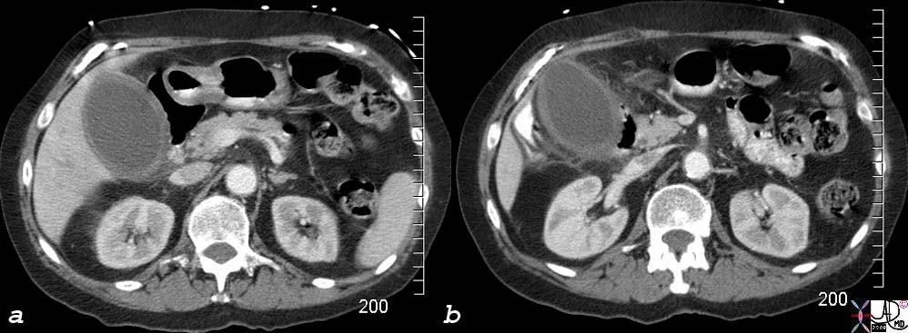
Normal or Large s/p ERCP? |
| 82141.8s gallbladder distended dilated s/p ERCP CTscan Courtesy Ashley Davidoff MD copyright 2008 |
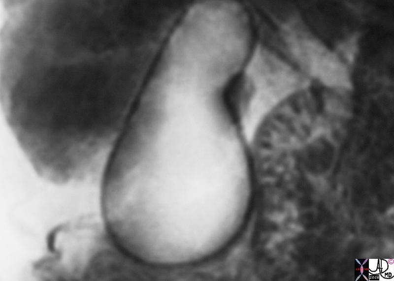
Pear Shaped Gallbladder |
| 17082b01.8s gallbladder distended MRI T2 weighted CBD stone choledocholithiasis Courtesy Ashley Davidoff MD copyright 2008 |
Acute Pancreatitis |
| 26898c code pancreas + peripancrteatic tissue + fx fluid accumulation + dx acute pancreatitis CT scan C+ code gallbladder |
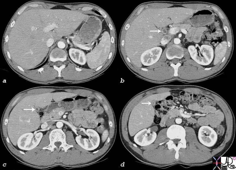
Sclerosing Cholangitis |
| 25187c.8s 29 female with obstructive jaundice bile ducts thickened heterogeneous ulcerative colitis sclerosing cholangitis gallbladder dilated fluid in gallbladder fossa distended CTscan Courtesy Ashley DAvidoff MD copyright 2008 |
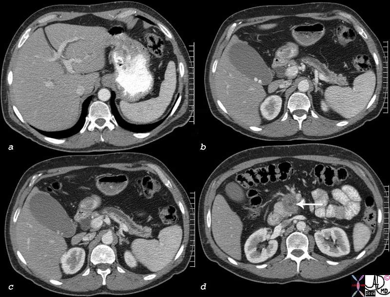
Courvoisier Gallbladder |
| 78441c01.8s painless jaundice pancreas mass bile ducts intrahepatic dilated extrahepatic gallbladder enlarged distended dilated gallstones cholelithiasis pancreatic duct double duct sign Courvoisier’s CTscan Courtesy Ashley Davidoff MD copyright 2008 |

Sclerosing Cholangitis |
| 04202 bile duct + sclerosing cholangitis ulcerative colitis gallbladder enlarged obstructed cholestasis |
Sclerosing Cholangitis |
| 27089b gallbladder + fx large + dx sclerosing cholangitis + imaging radiology MRI T2 + MRCP bile duct stenosis |
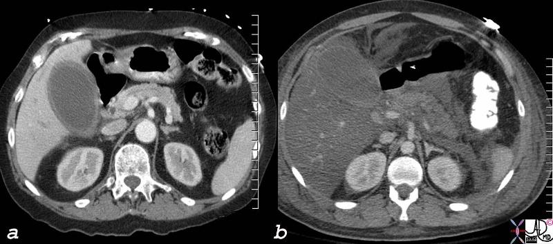
Dilated in Acalculous Cholecytitis and Dilated in Fatty Liver |
| 78606c.8s liver normal liver abnormal fatty change 777017 = a = acalculous cholecystitis b= pancreatitis with steatosis hepatization of the gallbladder density of the liver is the same as the bile in the gallbladder CTscan Courtesy Ashley Davidoff MD copyright 2008 |
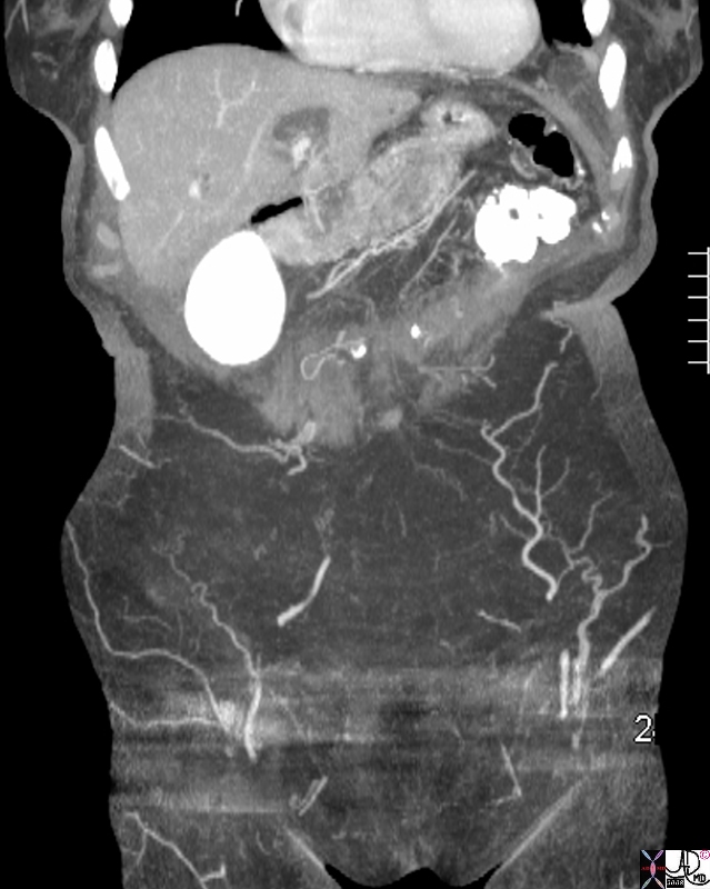
Extrinsic Impression by Dilayed Gallbladder on Bile Duct |
| 82150c01.8s bile duct extrinsic compression by enlarged gallbladder ERCP Courtesy Ashley DAvidoff MD copyright 2008 |
It is a blind sac with a muscular and elastic layer, giving it the ability to dilate when it contains bile and the ability to contract when it has to deliver bile to the duodenum.
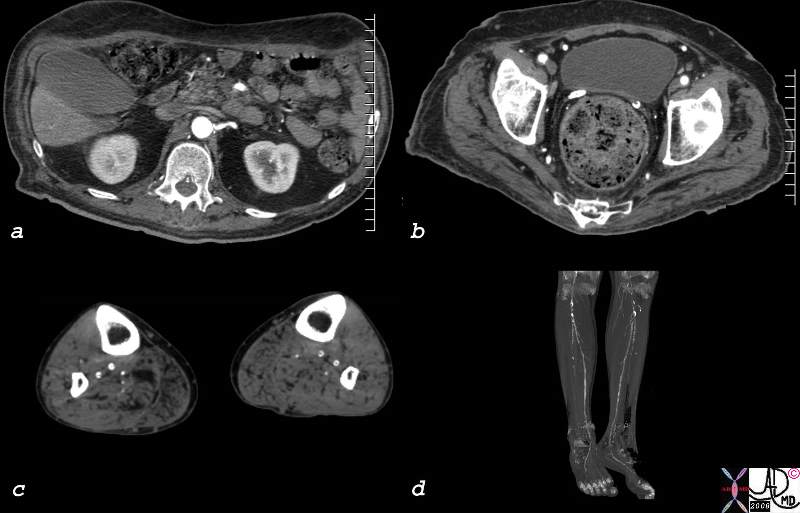
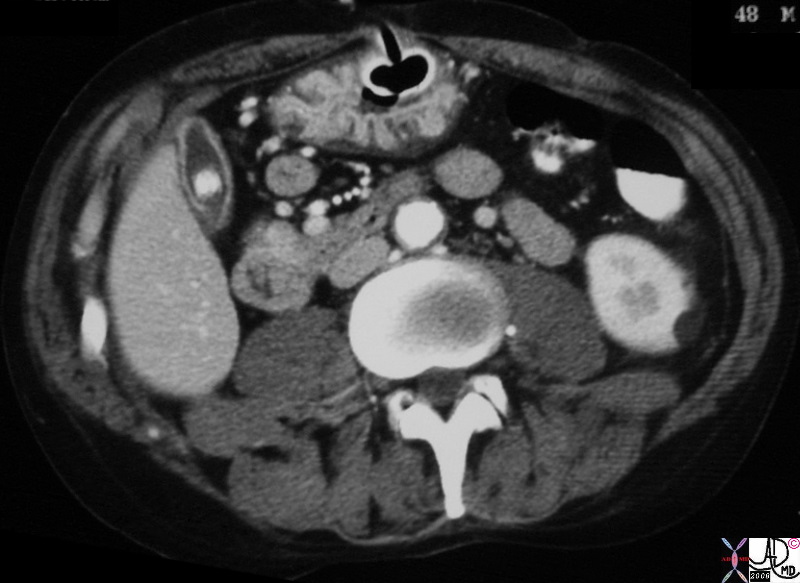
Diabetic Arteriopathy – Gallbladder Atony |
| 82719c.8s 68M diabetes peripheral vascular disease diabetic arteriopathy artery small vessel disease gallbladder atonic enlarged vas deferens calcification calcified Courtesy Ashley Davidoff MD copyright 2008 |
prolonged fasting also results in a large gallbaladder. When the transverse diameter exceeds 4cms it is abnormal but included in this differential is the fasting gallbladderlogical
Depending on the clinical presentation of the patient, the gallbladder may be more distended if the patient has not eaten for some time or contracted if he or she has recently eaten.
Distended Gallbladder |
| 00384 gallbladder pancreas liver anatomy |
References
Dodds, Wylie
Pathology Outlines: Gallbladder