
aka The Bare Area of the Gallbladder
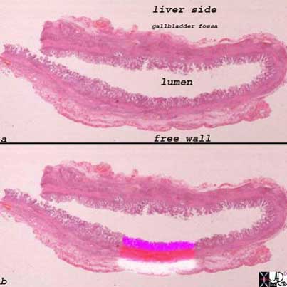
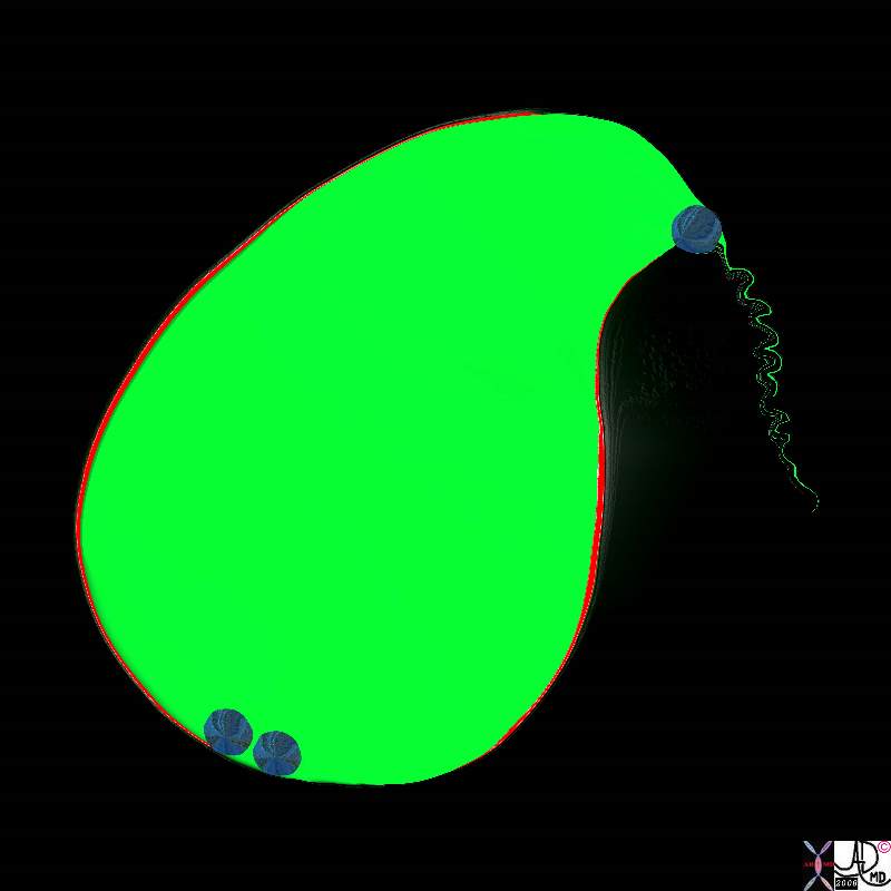
The upper and lower images represent whole mounts of a normal gallbladder. The upper image shows the liver surface of the gallbladder and the region of the gallbladder fossa, the lumen, and the free wall that is covered by the peritoneum.
The lower image shows the three basic histological layers. The pink mucosa, the red muscularis, and the white outer serosa.
Images courtesy of: Ashley Davidoff, M.D.2008
A closer look at the bare area of the Gallbladder
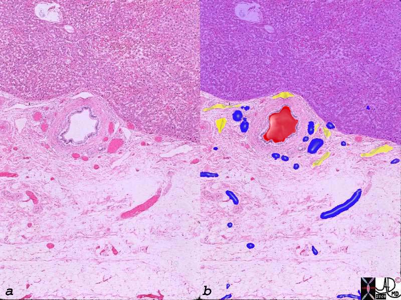
Arteries, Veins and Lymphatics in the Gallbladder Fossa |
|
The histological section of the gallbladder fossa shows the relatively large thick walled branch of the deep cystic artery, abutting the liver (upper portion purple) accompanied by venules (blue) and lymphatics (yellow). The veins and lymphatics drain directly into the liver. 00140c03.8s gallbladder wall gallbladder fossa normal artery duct liver interface histopathology |
The gallbladder fossa is rich in lymphatics
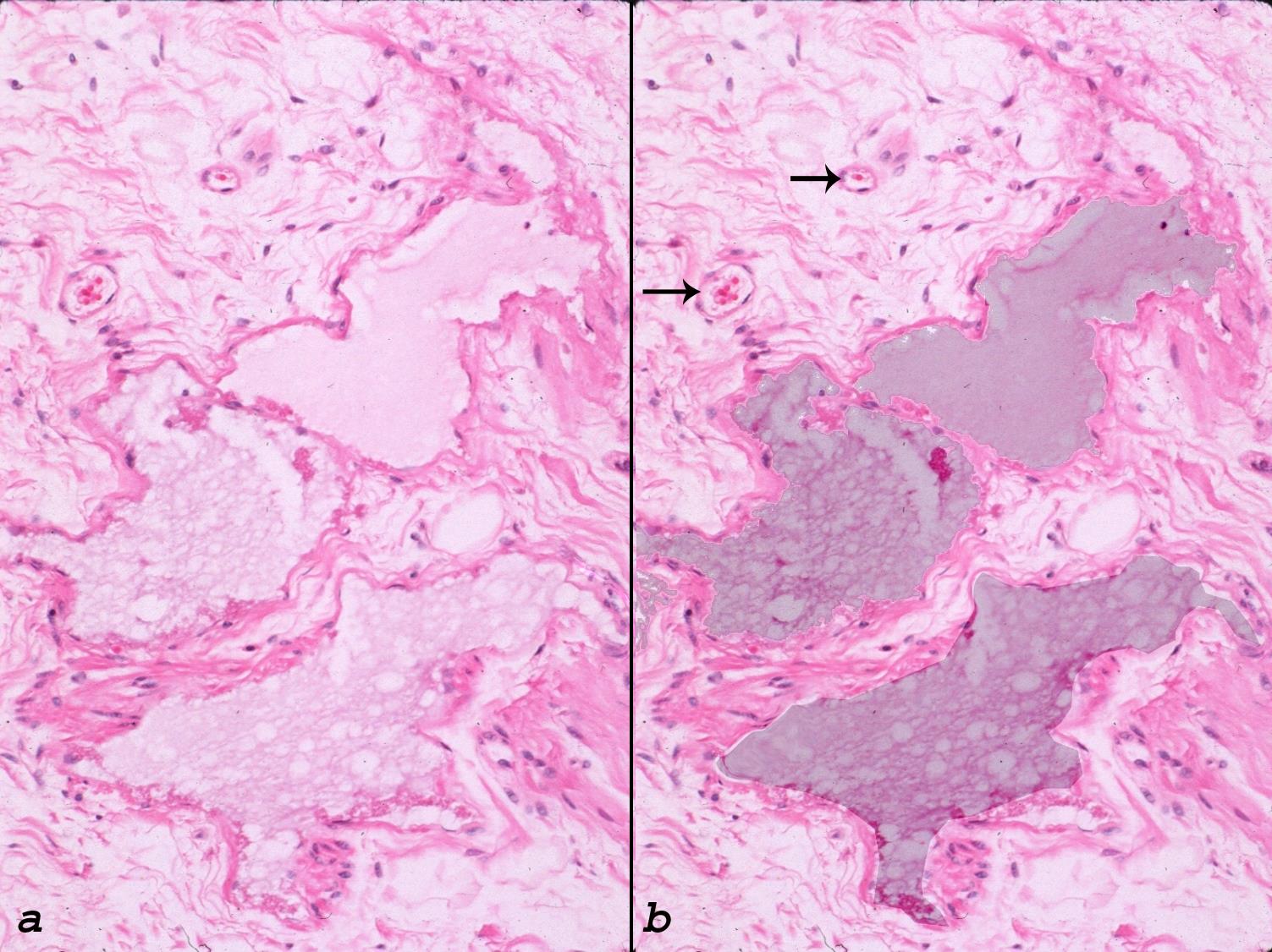
The gallbladder fossa is the hilum of the gallbladder and contains lymphatics, venules and arterioles and sometimes there is direct drainage of the biliary system into the gallbladder (duct of Luschka)
In Acute Cholecystitis
In acute cholecystitis there is hyperemia with increased blood flow, congested capillaries and increased leakage of fluids resulting in edema and increased lymphatic congestion , Since the histology of the bare area and free wall of the gallbladder are so different they have different imaging manifestation. As the inflammatory process progresses the veins and lymphatics become congested and since they are concentrated in the gallbladder fossa, fluid and fibrin start to accumulate in the fossa. The fluid may be seen as a hypoechoic region on US, while if fibrin predominates a lacy pattern is seen
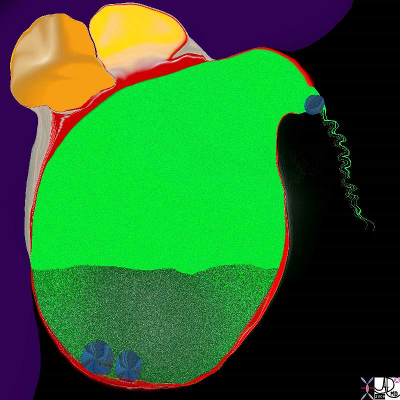
The longer the stone remains impacted, the more swollen the gallbladder becomes and the pressure on the wall causes ischemia and inflammation. The diagram shows a reddened and slightly thickened wall.
11921.8b05b020.8s gallbladder cystic duct gallstones cholelithiasis stone impacted in the cystic duct which is distended
Ashley Davidoff MD
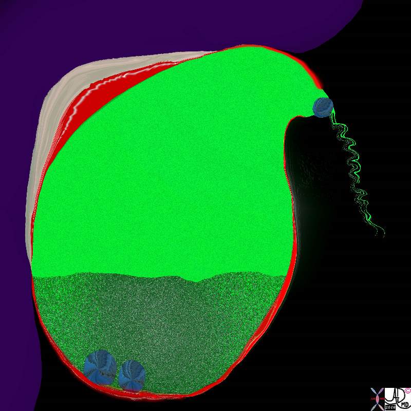
As the inflammatory process progresses the veins and lymphatics become congested and since they are concentrated in the gallbladder fossa, fluid and fibrin start to accumulate in the fossa. The diagram above accumulation of fluid (beige) and red cells (red) in the gallbladder fossa.
11921.8b05b031.8s gallbladder cystic duct gallstones cholelithiasis stone impacted in the cystic duct distended enlarged hyperemic wall complex fluid in the gallbladder fossa tumefactive bile cholestasis sludge acute cholecystitis Davidoff Art copyright 2008
Acute Calculous Cholecystitis with Gallbladder Wall Edema and Hyperemia |
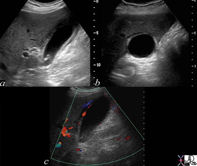
| The three images depicted above are from a patient with acute cholecystitis. Image a shows the gallbladder in longitudinal axis, with shadowing stones in the neck of the gallbladder, a thickened wall, and complex fluid in the gallbladder fossa. Image b shows the gallbladder in transverse view and again shows the complex fluid and soft tissue changes in the gallbladder fossa alongside the liver. Image c shows a longitudinal view of the gallbladder with hyperemia in the gallbladder wall abutting the gallbladder fossa, as depicted by color flow doppler.
72365c01 gallbladder wall fluid in gallbladder fossa positive sonographic Murphy’s sign inflammation mechanical obstruction infection hyperemia hypervascularity dx acute calculous cholecystitis Davidoff MD |
The accumulation of fluid in this area make blunt dissection for the surgeon relatively easy and surgeons say that if it is present they can use their fingers to dissect the gallbladder from the liver
Fluid and Thickening in the Gallbladder Fossa |
|
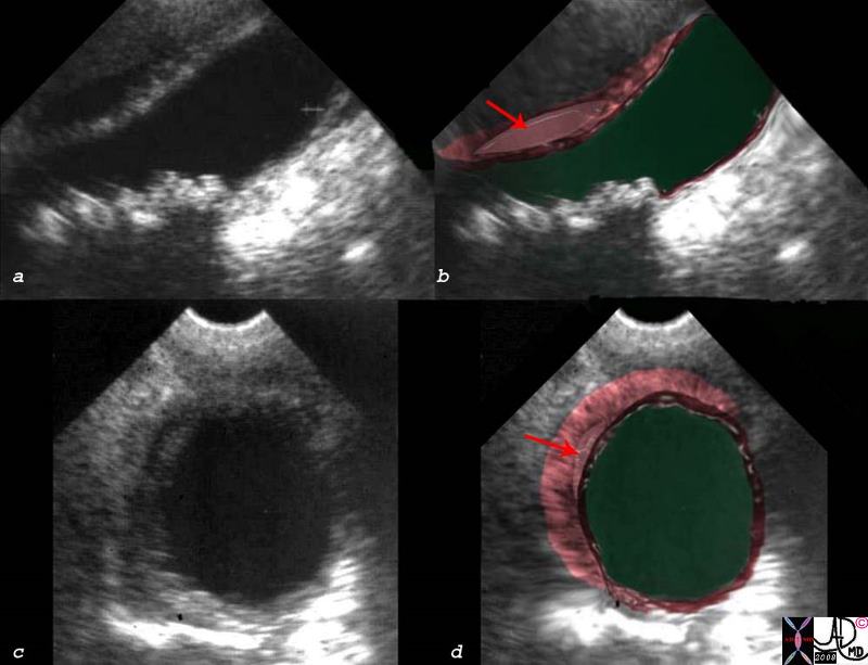
The patient presented with right upper quadrant pain RUQ pain, positive Murphy’s sign, fluid in the gallbladder fossa (red arrow in b and d)thickened linear lacy thickening (dark pink) and small shadowing stones (a,b) The diagnosis is consistent with acute cholecystitis. acute calculous cholecystitis USscan ultrasound Courtesy Ashley Davidoff MD copyright 2008 00543c03s.8 |
 Acute Cholecystitis00401.1cs 67Male right upper quadrant pain RUQ pain positive Murphy’s sign gallbladder gallbladder fossa thickened linear lacy thickening gall stones calculi calculous large stone in the infundibulum shadowing multiple small stones cholelithiasis cholecystitis acute cholecystitis acute calculous cholecystitis USscan ultrasound Courtesy Ashley Davidoff MD copyright 2008
 Acute Cholecystitis Caused by Cholesterol Crystals
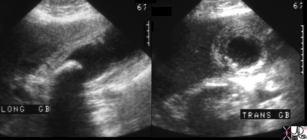
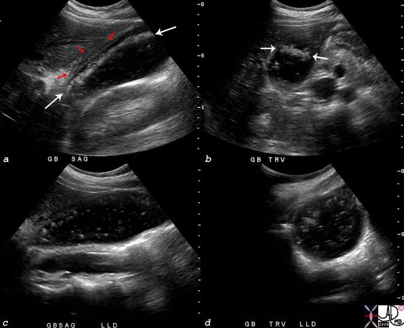
The ultrasound scan is from a young female who presented with right upper quadrant pain and positive Murphy’s sign – a clinical sign that is consistent with acute cholecystitis. The scan shows multiple floating stones (white arrows) in the longitudinal view (a) and in the transverse view (b). There are complex changes in the gallbladder fossa (red arrows) characterized by fluid and linear echoes, both characteristic of acute cholecystitis. Perturbation of the stones disturbed the layering effect and the crystals were seen floating throughout the lumen.
77753c03.8s young female right upper quadrant tenderness RUQ gallbladder echogenic irregular fluid fluid layer conforming to the shape of the gallbladder wall thickened linear echoes floaters crystalline SG less than bile cholesterol crystals specific gravity forces space cholelithiasis stones small acute cholecystitis USscan ultrasound Courtesy Ashley Davidoff MD For Radiologists and Detectives
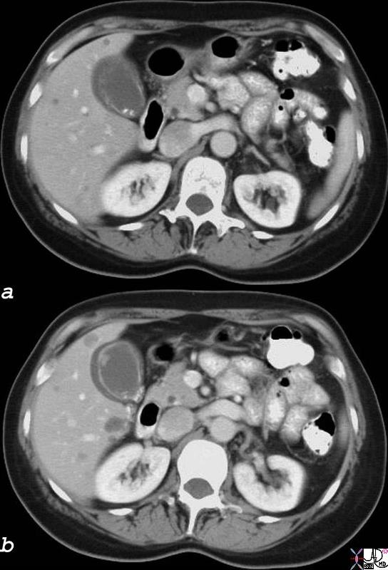 Acute Cholecystitis with Fluid in the Gallbladder Fossa 18333c01.8s gallbladder edematous wall hyperemic wall distended gall bladder edema in the wall fluid in the gallbladder fossa GBF fluid acute cholecystitis stones visible on CT scan cholelithiasis gallbladder polyp CTscan Courtesy Ashley Davidoff MD copyright 2008
Spread of Disease via the Gallbladder Fossa
If there is a superadded infection, spread to the gallbladder fossa and directly to the liver is the obvious route via the lymphatics and veins. The same holds true for the spread of gallbladder cancer – ie directly into the liver.
Eccentric thickening of the gall bladder fossa also occurs in patients with hepatitis, presumably due to increased lymphatic pressure in the liver as a result of the inflammation and thus impedance of lymphatic drainage from the gallbladder
Liver Abscess from Acute Cholecystitis
Cholecystitis Complicated by Abscess Formation in the Gallbladder Fossa
The artistic rendition shows 2 stones lying free in the lumen and one impacted in the neck of the gallbladder with secondary inflammatory changes in the wall(red) and abscess formation in the gallbladder fossa and extension into the neighbouring liver.
11921.8b05b037.8s gallbladder cystic duct gallstones cholelithiasis stone impacted in the cystic duct distended enlarged hyperemic wall complex fluid in the gallbladder fossa tumefactive bile cholestasis sludge acute cholecystitis abscess formation Davidoff Art copyright 2008
 Early Abscess Formation16188c05.8 gallbladder hyperemic thickened wall early abscess formation fluid collections in the gallbladder fossa acute cholecystitis CTscan Courtesy Ashley Davidoff MD copyright 2008
Spread of Cancer of the Gallbladder via the GBF to the Liver
Direct Invasion into the Liver and Bile Duct Obstruction
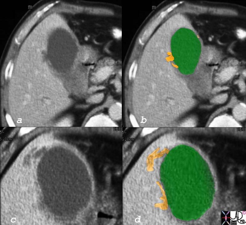
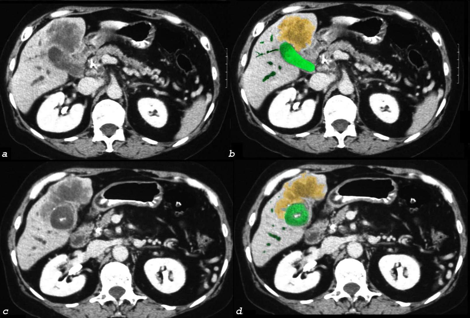
The CTscan of this patient shows a normal sized gallbladder (green) associated with a 4-5cms mass (orange) adjacent to the gallbladder, and extending from the gallbladder fossa. There is associated biliary obstruction (dark green tubes) This case represents invasive gallbladder carcinoma with bile duct obstruction. Images c and d show the almost universal association of gallstones in patients with carcinoma. In this case the stones (white) are in the centre of the gallbladder. (green)
16254c03.8s gallbladder anterior wall liver invasion space occupation obstruction bile ducts aggressive gallbladder carcinoma complicated by direct invasion metastasis liver windows narrow windows tumor settings CTscan copyright 2008
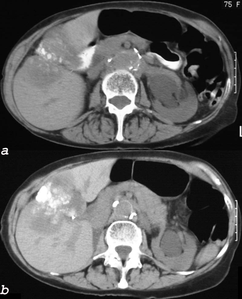
24404c.8s 75 female gallbladder calcification adjacent mass in the liver local invasion into the gallbladder fossa dystrophic calcification probably mucinous adenocarcinoma of the gallbladder carcinoma stones cholelithiasis hydronephrosis
GBF Enlargement in Hepatitis
In hepatitis fluid in the gallbladder fossa may also be present presumably due to lymphatic congestion in the liver and decreased lymphatic drainage from the gallbladder