
Copyright 2008
Alok Anand Ashley Davidoff MD
Definition
A hepatobiliary iminodiacetic scan, cholescintigraphy is an imaging procedure of the gallbladder that is used to evaluate the the gallbladder and biliary tree. Radioisotope is injected into the vein and is concentrated by the liver and excrted into the biliary system, and normally will be concentrated and visualized in the gallbladder and biliary system.
Principles
Technetium-labeled analogues of iminodiacetic acid (IDA) or diisopropyl IDA-DISIDA are injected intravenously. These substances are secreted by hepatocytes into bile. This enables visualization since as radioactive substances they emit , enabling visualization of the liver and biliary tree.
The radioactive isotopes are injected intravenously and are secreted by the hepatocytes into the biliary system. As the isotope decays, gamma rays are emitted, or “scintillates”. The scintillation is detected by a gamma camera or rectilinear scanner, both of which are cameras designed to detect light in a range of very high energy frequencies. On the images generated, “hot areas” represent a relatively higher uptake the radionuclide.
Technicium 99 is the most commonly used radioisotope. It has a half-life of 6 hours, and can be generated relatively easily using portable equipment that can be kept on premises of most imaging facilities. When imaging the gallbladder, Technicium 99 is usually attached to hepatobiliary iminodiacetic acid, which collects in the liver and is excreted into the bile ducts. It collects in the gallbladder before being expelled into the GI tract, and thus is useful for studying bile outflow. If after 4 hours post-injection a gallbladder does not fill , then obstruction is to be suspected.
Indications
The most common indication for cholescinitgraphy is in the case of suspected gallbladder obstruction. This is usually due to gallstones, but may also be due to space-occupying tumors. It can also be used to evaluate leaks of the biliary tree.
Contraindications
The only absolute contraindication to the use of HIDA scan is pregnancy, as the radionuclide may localize to the liver of the fetus.
Advantages
Advantages of scintigraphy is reduced radiation dose over CT. Additionally, HIDA scans are considered to be diagnostically more specidic and accurate than ultrasound for acute cholecystitis.
Disadvantages
This requires intravenous injection followed by a wait time of up to several hours for a definitely positive scan, although negative results can be obtained much sooner. Another disadvantage is that it does not clearly depict structural features of the gallbladder as well as ultrasound or CT. In these modalities, wall thickening can also be seen, which is helpful in the diagnosis of acute cholecystitis.
HIDA scan cannot be perfrmed in patients whose bilirubin >4.4mg/Dl
Aim
The aim of the study is to evaluate the function and patency of the gallbladder and biliary system.
Method
Patient Preparation
Patients are instructed to not eat or drink four hours prior to the exam. The patient is told to be prepared to be studied over a 4 hour period. The patient needs to lie still and supine during the acquisition of images
Technique
The radionuclide is injected intravenously, and the patient is placed under the gamma camera. A wait of up to fours may be necessary in cases to definitively diagnose obstruction.
Equipment
A gamma camera is neede to detect the radioactivity emitted.
Result
The gallbladder should be visualized within 30 minutes. The scan is considered positive if the gallbladder is nort visualized in 60minutes. Sometimes hyperemia of the gallbladder fosssa is seen and this is called the rim sign. Although not definitively known, the sensitivity and specificity of HIDA scans is approximately 97–100% and 93–95%, respectively
If the small bowel is not visualized in 60 minutes then obstruction of the bile duct is diagnosed.
If there is radioactivity outside the bowel then a leak of bile is confirmed.
False positives occur in patients with cholestasis and in these circumstances morphine is given to causethe sphincter of Oddi to close and is an attempt to force bile up the cuystic duct.
Examples of Various Diseases
| Positive HIDA scan |
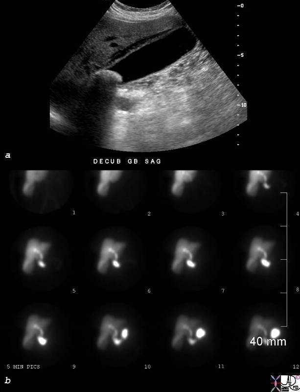
| 78306c01.8s young female vague right upper quadrant pain non distended gallbladder complex fluid in the gallbladder fossa large stone cholelithiasis non visualization non viz radioactive labelled technetium HIDA scan non visualization of the gallbladder at 45 minutes biliary system visualized at 30 minutes consistent with acute cholecystitis nuclear medicine Courtesy Ashley Davidoff MD copyright 2008 |
 Positive HIDA scan Positive HIDA scan |
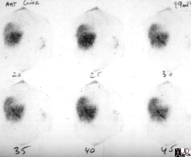
| 04505bs radioactive labelled technetium HIDA scan non visualization of the gallbladder at 45 minutes biliary system visualized at 30 minutes consistent with acute cholecystitis nuclear medicine Courtesy Ashley Davidoff MD copyright 2008 |
HIDA scan Positive for Acute Cholecystitis |
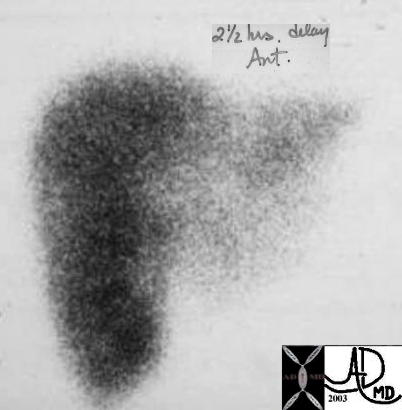
| The scan was taken at 2 and half hours after administration of the radiosotope, and fails to show filling of the gallbladder. This finding confirmins an obstruction of the cystic duct and the diagnosis of acute cholecystitis is highly likely.
04208 gallbladder non-filling HIDA scan dx acute cholecystitis imaging radiology nuclear medicine NMscan |
Complications
No complications are encountered with this study
Conclusion
The HIDA scan is the most sensitive study in the diagnosis of acute cholecystitis and for bile leaks. Howevere it is only usually used for cholecystitis in atypical cases when ultrasound findings are equivocal. False positives occur with cholestasis
References
Journal of Emergency Medicine – Diagnostic Utility of Cholescintigraphy in Emergency Department Patients with Suspected Acute Cholecystitis: Comparison with Bedside RUQ Ultrasonography
Ziessman, Harvey A.,. Fahey, Frederic H , and Hixson Donald J. Calculation of a Gallbladder Ejection Fraction: dvantage of Continuous Sincalide Infusion over the Three-Minute Infusion Method J NucIMed1992;33:537- 541