
Copyright 2008
Alok Ananad MS IV
Ashley Davidoff MD
Definition
Hydrops is a chronically distended gallbladder that contains mucous or serous type bile caused by chronic obstruction most commonly as a result of a chronically impacted stone.
Structurally it is distended, and contains relatively clear or mucoid secretions. This is usually in the setting of a solitary obstruction, usually at the gallbladder neck or in the cystic duct. It is usually non-inflammatory. The wall may be relatively thick or thin. In cases of chronic or recurrent obstruction, over time reabsorption of the bile and bile salts by the gallbladder mucosa occurs. The resultant clear or mucoid liquid, sometimes called white bile, is present.
Functionally there is no large consequence to overll body functions in this disease entity.
Clinically roughtly three percent of all cases of gallbladder pathology are concomitant with hydrops fo the gallbladder. In children it may be associated with with hemorrhagic dengue or even Kawasaki’s disease. (subcutaneous lymphatic syndrome).
The patient presents with RUQ pain, usually nonicteric, and in the absence of fevers or chills. A palpapable, slightly tender gallbladder is usually noted.
Laboratory and serology will not usually be abnormal, though may approach the upper limit of normal, particularly alkaline phosphatase and white cells, depending on the degree of inflammatory involvement.
Imaging diagnosis usually depends on ultrasonography. Non tender gallbladder distension with a stone in the neck of the gallbladder would be the classiocal presentataion. Wall thickening is usually present, often due to recurrent cholecystitis. Rokitansky-Aschoff sinuses, owing to the increased luminal pressure of the gallbladder may be seen. Though this syndrome is usually considered grossly non-inflammatory, on microscopic examination, white cells maybe noted interspersed amongst the flattened mucosal cells.
Definitive treatment requires cholecystectomy.
Chronic Impaction – Hydrops of the Gallbladder |
|
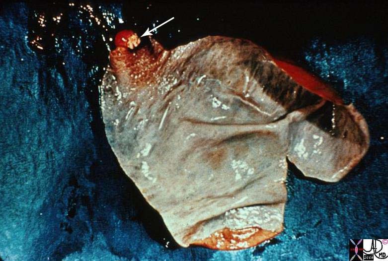
This is a gross photograph of a gallbladder opened longitudinally to show the mucosal surface. The gallbladder is distended, and its walls are very thin. Look carefully and note part of a light tan, granular gallstone protruding from the neck of this gallbladder, where it is impacted. This impaction has obstructed the gallbladder, preventing bile from getting in from the liver. The columnar cells lining the gallbladder keep producing mucin, their normal function, and the mucin cannot exit the gallbladder. Therefore, the gallbladder gets progressively distended with a clear, glairy mucin, and this situation is called hydrops of the gallbladder. 11917.8s01 gallbladder dx cholelithiasis grosspathology Courtesy Barbara Banner MD |
Hydrops of the Gallbladder |
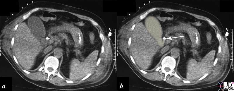
| The CTscan is from an elderly man who presented with mild right upper quadrant pain and a fever. There was no definite Murphy’s sign but there was definite mild discomfort to palpation. The scan shows a mildly distended gallbladder with a thickened wall and no pericholecystic fluid nor obvious induration of the fat. It does however show a calcification in the neck or cystic duct of the gallbladder (arrow) which reflects a chronically impacted stone. The gallbladder was drained percutaneously and the “bile” was almost like water in color, but with a slightly thickened texture. These findings are classical for hydrops of the gallbladder.
82335c01.8s gallbladder distendedd thick walled stone in the cystic duct pale yellow almost white fluid drainage clear fluid hydrops of the gallbladder CTscan Courtesy Ashley Davidoff MD Copyright 2008 percutaneous drainage cholecytostomy |