
The Common Vein Copyright 2008
Alok Anand Ashley Davidoff MD
Definition
MRI of the gallbladder is an imaging study, usually utilised if other studies such as ultrasound and CT imaging have been unyielding or difficult to perform. MRI is highly sensitive to water and hence the gallbladder and its surrounding structures and biliary system are exquisitely visualized.
|
|
Principles
Magnetic Resonance imaging, unlike other forms of imaging, does not utilize ionizing radiation from accelerators or radioactive sources. Instead, it utilizes the uniqe magnetic properties of matter in the body to construct anatomical images. Hydrogen, which is present in nearly all compounds of the body, has the ability to act like a magnet. In the presence of a relatively weak field, such as that created by the earth the alignment of the atoms of hydrogen are randomly assorted throughout the body. In the presence of stronger magnetic fields, however, these particles start to line up, like the needle of a compass. Once aligned, these atoms are excited with a radio frequency light pulse. When excited, these atoms can release a pulse of their own. The type of energy depends on several factors:
-The strength of the magnetic field
-The bonding relationship between the hydrogen atoms and nearby atoms.
Sophisticated imaging equipment uses these two pieces of information to analyse the signals emanating from the body to construct a picture of the tissues within.
Indications
MRI is highly sensitive to water and hence the gallbladder and its surrounding structures and biliary system are exquisitely visualized.
Contraindications
The intense magnetic fields of the MRI magnet can easily interfere with electronics or magnetic materials. As such, the use of MRI is contraindicated in individuals with unremovable electronic devices, such as pacemakers. Although most implants and prostheses manufactured today are MRI safe (see: http://www.mrisafety.com/), caution should be exercised when anyone with metallic objects in their body are suspected, and verifications of these materials is a must. In addition, free metals may be present in the body, such as metal filings in the eye. These objects under a magnetic field may move and cause damage to nearby soft-tissue structures.
Advantages
MRI can provide extremely detailed images of soft tissues. In addition techniques for rapid image acquisition have allowed use of MRI in many different settings, and is able to particpate in functional studies as well. MRI provides outstanding contrast resolution.
Disadvantages
MR images take considerably longer than other modalities of tomography. In addition, the bore magnets used tend to be small, and often cause claustrophobia in patients
Aim
The purpose of the study is to outline morphological features of the gallbladder and bile ducts and to define pathological entities
Method
Patient Preparation
Patient should be fasting and information regarding the length of the study, and should receive an explanation of what to expect during the procedure. Making the patient both physically and emotionally are helpful top the outcome of the examination since any movement for example will degrade the study.
Technique
The patient is place supine on the couch of the MRI within the bore of a magnet, and coil is appropriately positioned to generate the necessary radio pulse sequences.
Equipment
The equipment requires three elements; a static magnetic field, a radiofrequency transmitter and receiver and three magnetic gradients that can be controlled and are orthogonal to each other. The magnet is the largest component and form a doughnut around the patient. permanent magnets, resistive magnets, and superconducting magnets have been used. The higher the the field strength the greater the signal to noise ratio, but there are safety considerations in the magnets with high fild strengths.
Phased array coils use a paralel imaging technique to acquire multiple channels of data
The gradient coils are used to spatially encode the data acquired
Technique
The technique utilised for studying the gallbladder usually requires a T1 weighted sequence, T2 weighted sequence and gadolinium enhanced sequence.
Result
On T1-weighted images, the gallbladder wall has intermediate signal intensity and enhances uniformly after the administration of gadolinium-based contrast material. The portion of the gallbladder wall adjacent to the liver may not be well appreciated owing to similar enhancement of the wall and the liver parenchyma. On T2-weighted images, the gallbladder wall has low signal intensity and stands out against the bright visceral fat. The wall adjacent to the liver cannot be identified as a separate structure.
Normal bile appears uniformly bright with T2-weighted sequences. On T1-weighted images, bile varies greatly in signal intensitydepending on its concentration.
Gallbladder sludge may have similar signal intensity characteristics, namely, iso- to mild hyperintensity on T2-weighted images andhyperintensity on T1-weighted imagesThe sensitivity and specificity of MRI in diagnosing gallbladder pathologies depends heavily on the suspected pathology.
Examples of Various Diseases
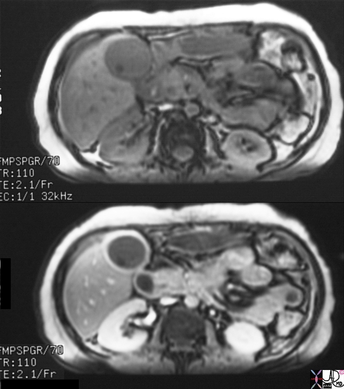
 Gallbladder Distension Associated with Common Bile Duct Stone Gallbladder Distension Associated with Common Bile Duct Stone |
| 17089c01.8s gallbladder enlarged choledocholithiasis bile duct obstructed fluid fluid level MRIscan T2 weighted T1 weighted Courtesy Ashley Davidoff MD copyright 2008 |
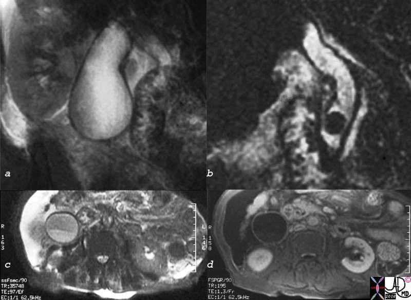
Acute Cholecystitis |
| 16141c01s.8 gallbladder stones cholelithiasis hyperemic wall edematous wall small filling defects in dependant position in the infundibulum distended gall bladder edema in the wall fluid in the gallbladder fossa gbf normal bile duct MRI T1 weighted image with gadolinium and fat saturation T2 weighted MRCP normal pancreatic duct Courtesy Ashley Davidoff MD copyright 2008 |
|
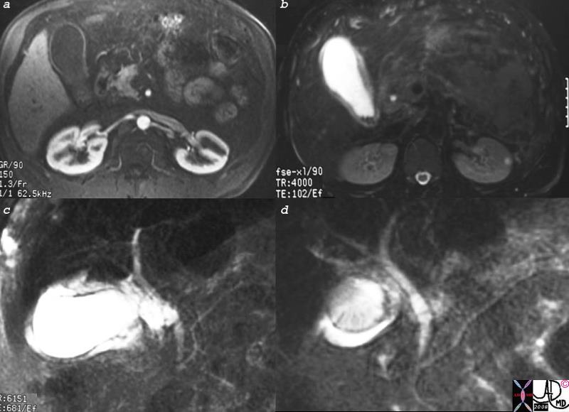
Acute Cholecystitis |
| 16141c01s.8 gallbladder stones cholelithiasis hyperemic wall edematous wall small filling defects in dependant position in the infundibulum distended gall bladder edema in the wall fluid in the gallbladder fossa gbf normal bile duct MRI T1 weighted image with gadolinium and fat saturation T2 weighted MRCP normal pancreatic duct Courtesy Ashley Davidoff MD copyright 2008 |
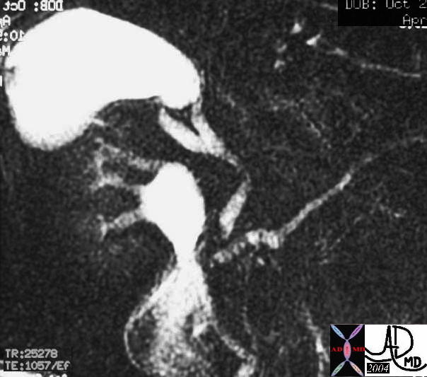
Distended Gallbladder Due to Sclerosing Cholangitis |
| 27089b gallbladder + fx large + dx sclerosing cholangitis + imaging radiology MRI T2 + MRCP bile duct stenosis |
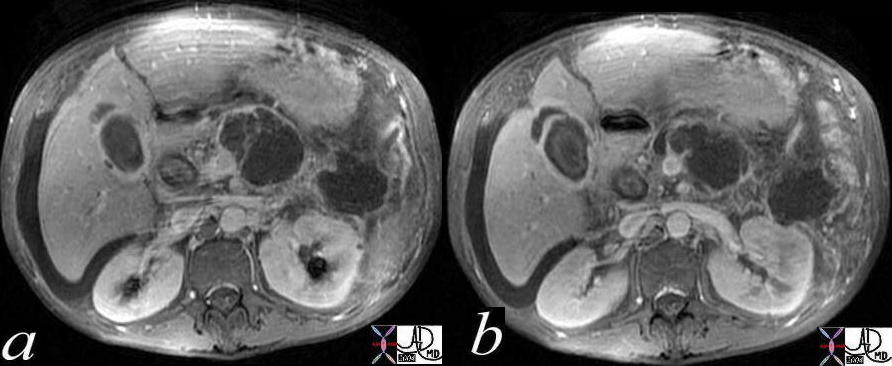
HIV Cholangiopathy |
| 41267c01 Courtesy Ashley Davidoff MD code pancreas pseudocysts code gallbladder fx fluid in the fossa dx HIV cholangiopathy chronic pancreatitis imaging radiology CTscan inflammation infection |
Chronic Cholecystitis Enhancing Wall |
| 16239c.8s gallbladder stone cholelithiasis wall thickened thick wall enhancing wall dx chronic cholecystitis bile duct cholangiocarcinoma MRI with and without contrast |
Complications
The
Conclusion
The
References
Name text publisher
Name :Article title journal volume year pages
Web References
MRI is useful in imaging the structures of various tissues. A combination of techniques, as well as the use of contrast agents to highlight different structural and chemical characteristics. The different protocols allow greater differentiation of soft tissue structures, thus cartilaginous areas, neural and soft tissues are particularly well imaged. In addition, by virtue of its design, MRI can be used to directly image in any plane or orientation.
MRI is commonly used in the gallbladder to diagnose carcinoma, as well as calculi within the gallbladder, as well as pathologies of the biliary tract, including sclerosing cholangitis, and HIV cholangiopathy. It can also distinguishes between the changes of the acute and chronic cholecystitides.