
Capsule of the Gallbladder
Copyright 2008
Definition
The gallbladder capsule is a protective covering consisting of loose connective tissue.
Structurally the coverings on the free surface and the hepatic surface are different. On the free surface the loose connective tissue is covered and protected by the peritoneum and it is called a serosa.. On the liver surface it is covered and protected by the liver and here it is called an adventitia. The fundus is totally surrounded by peritoneum. Occasionally the peritoneum almost totally surrounds the body of the gallbladder as well such that if one would place ones thumb over the anterior surface and ones index posteriorly they would meet separated only by a double layer of peritoneum. Usually though the gallbladder is bound down by the peritoneum so it remains snug and protected.
Fuctionally the serous membrane formed by visceral peritoneum covering is resilient, smooth and is richly endowed with nerves and lymphatics. It protects as well as connets the gallbladder with the other peritoneal covered organs of the abdomen. The adventitia acts more as aconduit to transmit fluids to and frm the mother organ the liver. It in fact is the hilum of the gallbladder.
The diseases of the coverings are really a mirror of disease in the peritoneal cavity, from withinn the gallbladder or from the liver. Serosal implants from peritoneal carcinomatosis, spread of inflammation from or to the liver, and spread of cancer of the gallbladder via the gallbladder fossa to the liver are some examples.
Treatments of the primary disease will therefore affect the way the capsule is treated.
Serosal Surface |
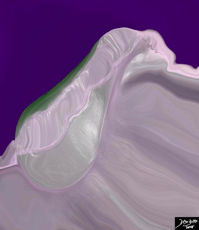
| The serosal surface of the gallbladder is covered by visceral peritoneum which then continues over the liver surface. the diagram shows the visceral peritoneum peeled back off the liver to exposed the bare are of the gallbladder which is really its adventitial surface, and is synonymous with the gallbladder fossa and bare area of the gallbladder.
04766b05b04.52k.8s gallbladder support protection peritoneum bare area gallbladder fossa normal anatomy gastrohepatic ligament peritoneum serosal surface adventitial surface Davidoff art copyright 2008 |
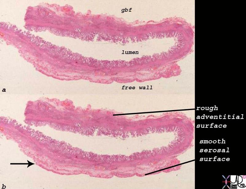
The histological sections seen below show the rough liver edge and the mesothelial covered free surface which is smooth
Whole Mount |
| When the two surfaces of the gallbladder are reviewed histologically on the whole mount, one sees the mesothelium (arrow) – a single celled layer of peritoneum that defines the smooth and glistening appearance of the free surface. Larger blood vessels can be seen on the raw surface of the gallbladder – the adventitia.
00139d.8s gallbladder histology normal gallbladder fossa free wall peritoneal lining whole mount mucosa histology Courtesy Ashley Davidoff MD copyright 2008 |
The following images of the gallbladder in longitudinal and transverse dimension show from an imaging perspective haw the visceral peritoneum encloses the liver , covers the fundus of the gallbladder, spreads over the body of the gallbladder, and then encloses the structures of the porta hepatis.
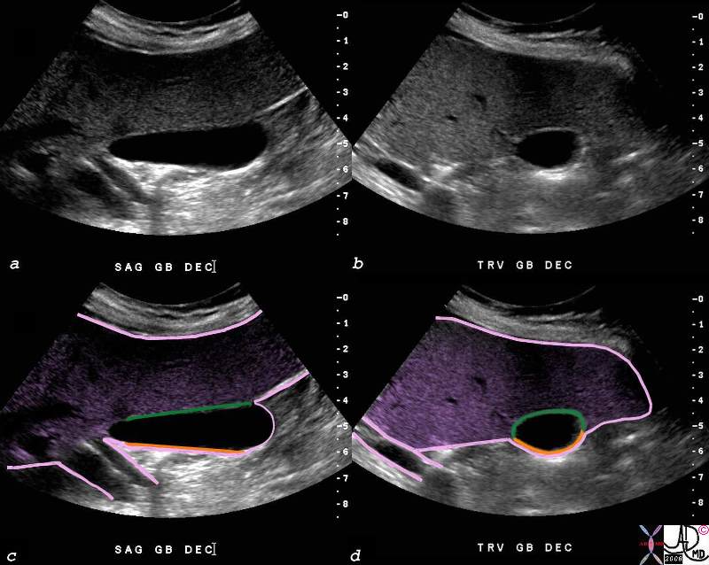
Peritoneal Covering of the Gallbladder |
| The visceral peritoneum (pink) encloses almost the entire liver, totally covers the fundus, of the gallladder as seen in the longitudinal view of the galbladder (c), and then covers the free wall of the body of the gallbladder on the undersurface of the liver (c,d) after which it reflects off the liver to enclose the the vessels of the porta hepatis Two tubular structures in the porta are surrounded by the same peritoneal lining.
82428c06.8s gallbladder small normal transverse oval shape gallbladder fossa Glisson’s capsule gastrohepatic ligament normal anatomy body of the gallbladder green is wall attached to the liver orange is free wall peritoneum = pink serosa liver capsule USscan ultrasound copyright 2008 Courtesy Ashley DAvidoff MD |
Applied Biology
When there is peritoneal carcinomatosis metastattic disease is able to settle and grow on the gallbladder. In the case shown below a metastsasis is seen in the fundus of the gallbladder.
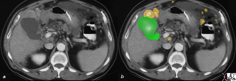
Metastases to the Fundus of the Gallbladder |
| The CTscan in cross section shows a deformed fundus of the gallbladder (green) compressed by a metastasis (orange) in this patient who has peritoneal carcinomatosis. Other metastases are noted in the peritoneal cavity and are also overlaid in orange.
22704c01.8s gallbladder abdomen peritoneum peritoneal cavity fx mass fx calcification fx distortion dx metastatic colon carcinoma large bowel malignant primary compliacated by transperitoneal spread dx metastasis to the gallbladder fundus metastases metastattic fx ascites imaging radiology CTscan C+ CTscan Courtesy Ashley Davidoff MD copyright 2008 |