
The Sac and the Wall
The gallbladder is a dark-green sturdy sac that looks like a deflated balloon when viewed anatomically. It has a soft leather feel due to its fibroelastic muscular wall and in essence can be described as distensible, muscular, and elastic.
Post Mortem Gallbladder Looks Like a Deflated Baloon. |
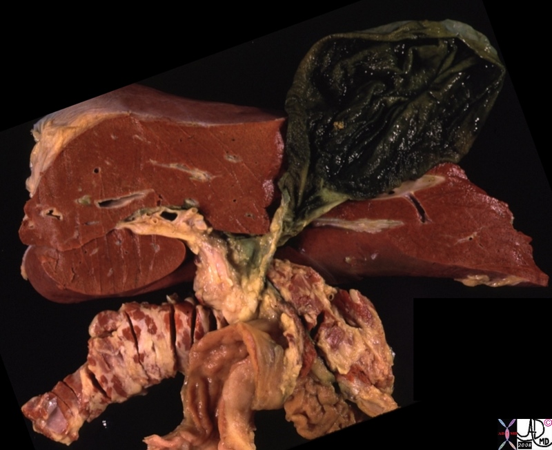
| 00384b.8s gallbladder dilated right lobe left lobe porta hepatis Glisson’s capsule pancreas liver normal anatomy grosspathology Courtesy Ashley DAvidoff MD copyright 2008 |
It is often stained with bile and crystalline granules. Once these are washed away the mucosa is usually pink and smooth. When the wall is cut in cross section the 3 layers are too thin to be distinguished macroscopically but three obvious layers are present histologically. The epithelium is cellular and the lamina propria as part of the mucosa is probably the loosest layer of the wall . The muscularis provides the musculo-fibro-elastic properties, while the serosa/adventitia also has releatively loose consistency.
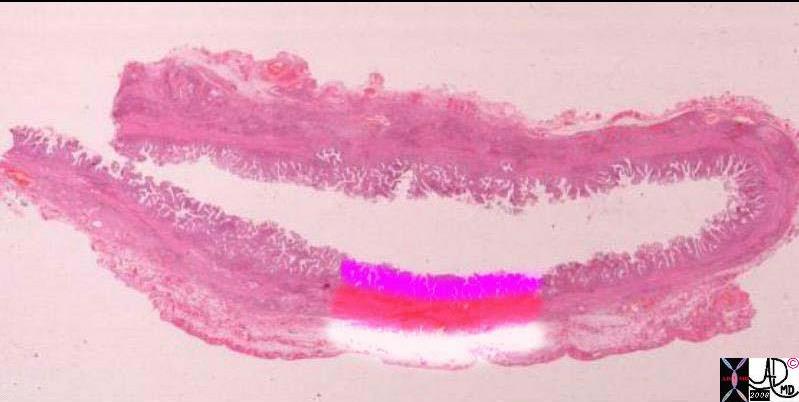
Character of the Wall is Based on the Character of Component Parts |
| The character of the gallbladder wall is based on the character of its component parts. Thus the epithelium is single cell layer thick . The lamina propria, which is part of the mucosa, (pink) is the loosest part of the wall. The muscularis layer (red) gives the gallbladder its musculo-fibro-elastic properties, while the serosa/adventitia (white) are loose tissues as well.
00139c11.81s whole mounts of a normal gallbladder. The upper image shows 3 relatively indistinct layers. The irregular luminal surface shows the folds of the mucosa under which the lamina propria runs (not obvious at this magnification). The layer below is the muscularis which is an poorly defined layer that consists of smooth muscle and connective tissue. (solid pink) The outer layer on the free wall (light pink) is covered by peritoneum and hence it is called a serosa. The inner layer lacks a serosal covering and hence it is called an adventitia. mucosa epithelium lamina propria loose muscular membranous elastic gallbladder histology normal gallbladder fossa free wall peritoneal lining whole mount mucosa histology Courtesy Ashley Davidoff MD copyright 2008 |
Bile
The bile is a bitter yellow or green alkaline fluid. It contains a variable concentration of solutes and water that change its character and specific gravity which ranges between 1.010 to 1.040 or even higher. It has a freezing point of -.56 degrees centigrade.
As it enters the cytic duct as fresh bile it’s SG is closer to 1.010. When it enters the neck mixes with mucus that is secreted in the neck resulting in a rise of SG . The gallbladder concentrates the bile by 5-6 times, eventually giving it an SG of about 1.040.
The principal constituents include salts of bile acids, bile pigments, neutral fats, lecithins, phosphatides, nucleoproteins, mucin and cholesterol.
Bilirubin gives bile its yellow-green color. It is the same pigment that gives the color to bruises and accounts for the yellow discoloration of jaundice. It is formed when hemoglobin breaks down.
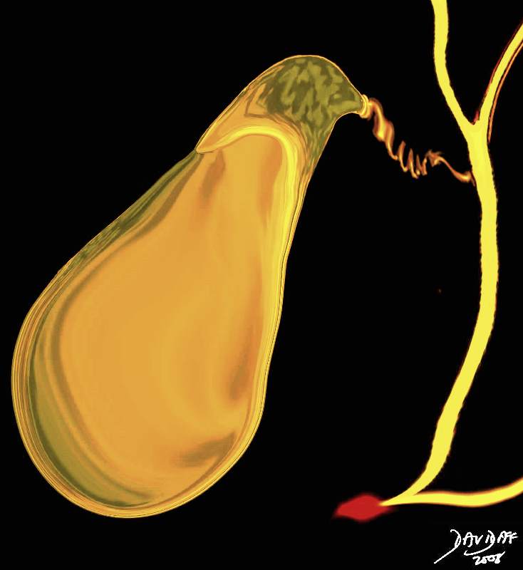
Yellow/Green Color of Bile |
| 04766b05b04.32k.8s gallbladder cystic duct right hepatic duct left hepatic duct common hepatic duct common bile duct pancreatic duct ampulla sphincter of Oddi normal anatomy Davidoff art copyright 2008 |
Applied Biology
We approach the characterization of the gallbladder in the clinical realm using a limited clinical exam, blood tests to assess and classify jaundice and medical imaging. From the structural perspective ultrasound is the best tool to characterize the wall and contents of the gallbladder.
When the normal gallbladder wall is imaged by ultrasound in the fasting state it stratified into its three layers. However when it is in its contracted, three definite layers are seen. The inner mucosal layer is echogenic, middle muscularis layer is hypoechoic and the outer serosal/adventitial layer is hyperechoic.
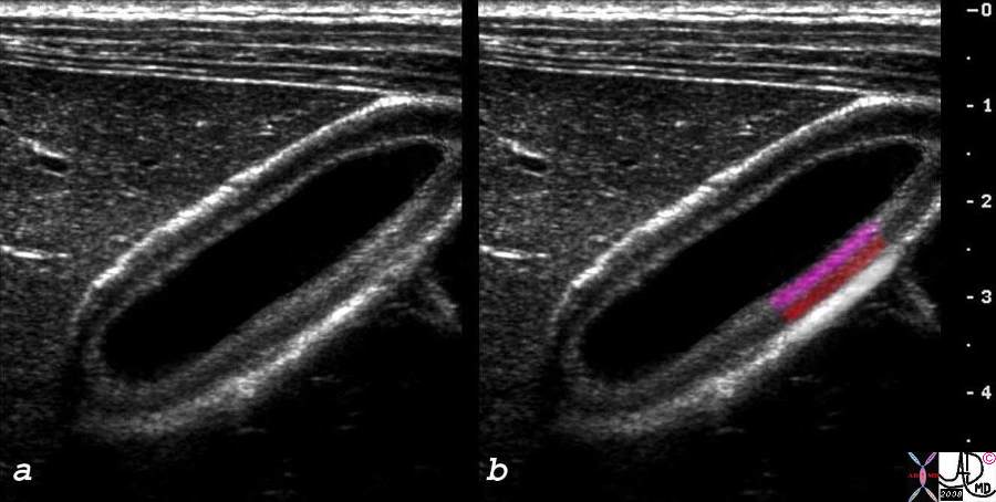
Characterization of the Wall by Ultrasound |
| The layers of the wall are only seen whenthe gallbladder is contracted or thickened for any reason. The inner pink layer is the mucosa, the miidle muscularis (red) and outer serosa/adventitial layer.
1632H5~1.9cs gallbladder contracted 3 layers mucosa muscularis serosa adventitia normal anatomy histology parts character size USscan ultrasound courtesy Philips Medical Systems Courtesy Ashley DAvidoff MD copyright 2008 |
There are many causes for an abnormal gallbladder wall but the example below shows a gallblader taken in the fasting state with a thick wall, where the inner layer is defined, the middle is thickened and outer layer is indistinct.

Thick Wall Normal Lumen – Hepatitis |
|
The three layers of the gallbladder are present but not normal and not well defined in this patient with hepatitis. The edema in the wall presumably pervades the loosest layers, and so it is assumed that the lamina propria and serosa/adventitia are mostly involved. The character of the lumenal content is still normal, but for a patient who has been fasting it is smaller than expected. 48008c01 gallbladder thick wall non distended dx hepatitis USscan Davidoff MD |
The bile containing lumen is anechoic in both the fasting state and postprandially.

Gallbladder in Fasting State Wall is a Single Layer and Bile is Anechoic -Clear Fluid |
| The sagittal view of the gallbladder shows a distended fundus and body of the gallbladder where contents have no echoes – clear fluid, and the only visible part of the wall abutting the liver is a single layer.
47018 gallbladder normal shape pear shaped anatomy nrmal USscan Davidoff MD |
The most common abnormalitis in the lumen include gallstones and sludge
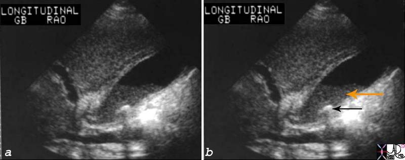
Stone Sludge and Tickened Gallbladder Wall |
| The sagittal view of the gallbladder shows a fluid-fluid layer with sludge (orange arrow) noted in the dependant portion, anechoic bile in the upper portion as the supernatant, and a shadowing stone (black arrow) lying dependantly in the body.
04740c01.8s gallbladder cholestasis fluid fluid level cholelithiasis shadowing stone thickening of gallbladder fossa GBF USscan Ultrasound Courtesy Ashley Davidoff MD copyright 2008 |
Advanced Knowledge on the characterization using imaging techniques can be found in the “character” section of imaging
References
Block The Practice Of Ultrasound: A Step-By-Step Guide to Abdominal Scanning By Berthold Block Published by Thieme, 2004
Hsu-Chong et al Floating Gallstones in Bile without Added Contrast AJR 1986
Tera H. Stratification of Human Gallbladder Bile In Vivo Acta Chirg Scan (Suppl) 1960 256: 9-85 (not available Pubmed)
Hawk Practical Physiological Chemistry By Philip Bovier Hawk, Olaf Bergeim Published by Blakiston, 1918