
Introduction
The gallbladder, like every organ in the body consists of its unique parts, a capsule, and its connection to other parts of the body, much in the way a house or building consists of its component parts, an outer covering and cable,electrical and sewer lines that connect it to the other parts of the community.
The gallbladder as a one compartement house consists of a fundus, body neck, its northern wall is protected by the liver, while its walls on the south, east and west side are covered by the visceral peritoneum. Its connections to the system at large are via the cystic duct, cystic arteries and veins, autonomic and hepatic lymphatic systems.
The House and its Single Room
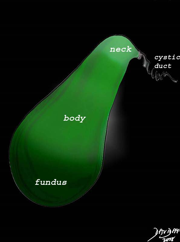
The gallbladder is divided into three general parts: fundus, body and neck. They are each very different in size shape and position, all relevant to the function of the gallbladder. The infundibulum of the gallbladder is part of the body and Hartmann’s pouch is part of the neck.
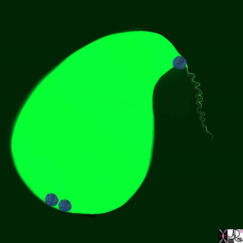
Parts of the Gallbladder |
| 04766b05b10.2k.81s gallbladder parts fundus = dark green body = green neck = light green normal anatomy
Davidoff Art Copyright 2008 |
Fundus
The fundus has the largest diameter, is usually spherical in shape, is the only portion of the gallbladder that is exposed and unprotected by the liver. It lies as the most anterior and rightward portion of the gallbladder. It lies superior to the hepatic flexure. As the most inferior portion of the gallbladder it is the most dependant part during the day while the person is upright. Hence the bile that is most concentrated with the highest SG will be found in the fundus. The blood supply is endarterial meaning it is the last part of the gallbladder to receive its blood and therefore it is most vulnerable to ischemia. It is the only portion of the gallbladder that is completely surrounded by peritoneum. Since it is not attached to the liver it is relatively mobile so that when the gallbladder stretches in atonic conditions such as old age, it is mostly the fundus that stretches.
 The Fundus as the Only Exposed Portion of the Gallbladder The Fundus as the Only Exposed Portion of the Gallbladder
Anterior Coronal View of the Gallbladder – MRI |
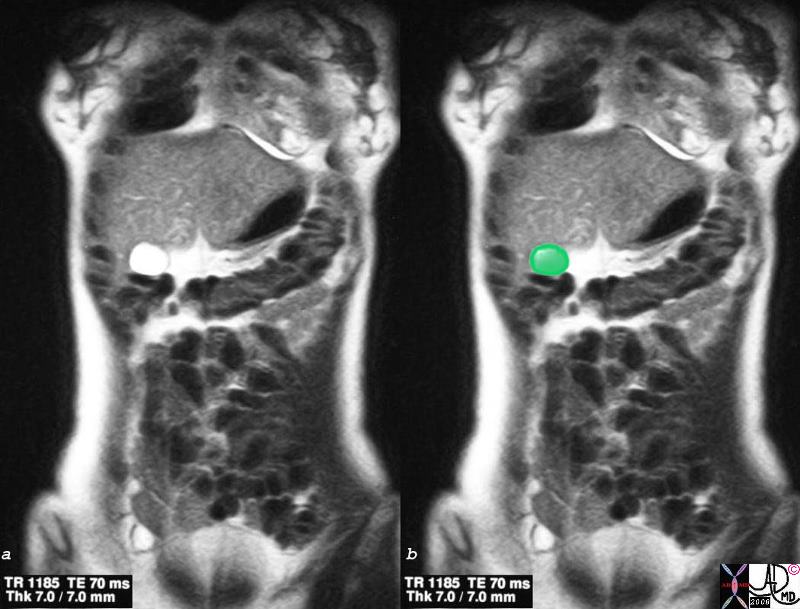
| Just beyond the liver edge in the midclavicular line, and just below the 9th right rib, the fundus of the gallbladder lies unprotected and potentially palpable to the examining hand. However it is not normally palpable unless it is significantly distended. The body and neck lie more posterior and inaccessible to clinical examination even when enlarged.
26629c.8s gallbladder liver normal anatomy fundus exposed anterior cut MRI T2 weighted Courtesy Ashley Davidoff MD copyright 2008 |
Applied Biology
The fundus is the only part of the gallbladder that potentially is clinically palpable, and in fact is only palpable when the gallbladder is significantly distended. When the pressure in the gallbladder rises the tension on the fundus will be maximal according to the law of Laplace since the tension on the wall for a given pressure is directly proportional to radius. Therefore the fundus with the largest radius will enlarge out of proportion in relation to the body and the neck. The site of rupture from distension therefore will also be the fundus not only because it has the most tension exerted on it but also because it is supplied by an end artery.
 Fundal Enlargement Fundal Enlargement |
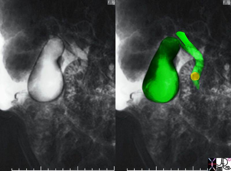
| The MRCP performed using T2 weighted FSE technique shows a CBD stone associated with dilatation of the CBD, cystic duct and the gallbladder. The shape change of the enlarged gallbladder is most profound in the fundus because of the increasesd tension exerted on the largest diameter of cylinder/sphere according too Laplace’s law.
17083c02.3k.8s 82 female presents with obstructive jaundice gallbladder duct enlarged distended distension dilated dilatation choledocholithiasis stone obstruction obstructive jaundice MRI T2 weighted Courtesy Ashley Davidoff MD copyright 2008 |
Body
The body of the gallbladder is the most voluminous portion of the gallbladder. It is more cylindrical in shape but its diameter narrows minimally as it advances from the fundus to the neck. The most medial portion of the body lies opposite the fundus, is tunnel shaped and is called the infundibulum.
The body of the gallbladder lies posterior to the liver and hence is relatively protected. The upper surface of the body lies in the gallbladder fossa and is free of a peritoneal covering. As a result of this attachment to the liver, the body is relatively fixed in position. The free surface of the body is covered by peritoneum and lies superior to and in close association with the duodenum. The muscular layer is best developed in the body of the gallbladder.
Body of the Gallbladder |
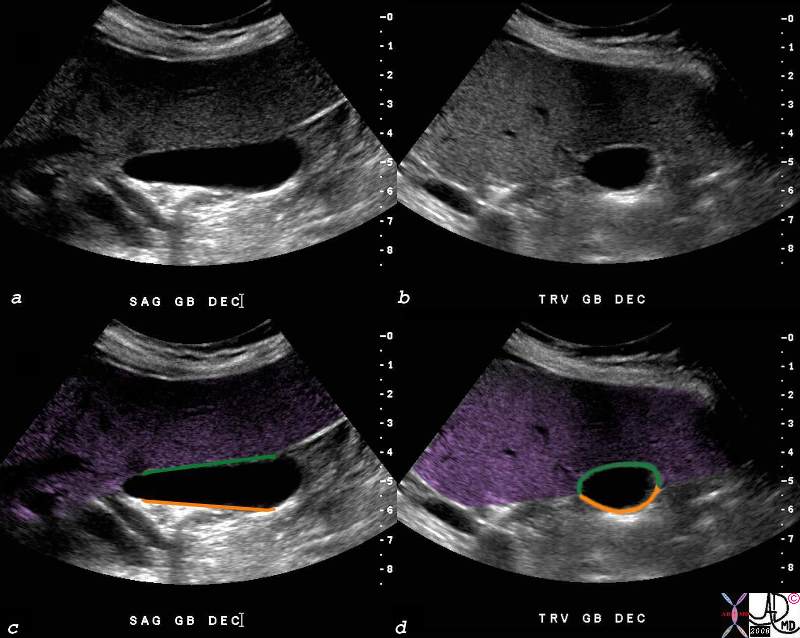
| The body of the gallbladder is attached to the liver.(purple) The US in sagittal section (a,c) and transverse section (b,d) shows the fixed portion (green) of the body attached to the gallbladder fossa and liver, and the free wall (orange) lying open to the peritoneal cavity. The image also reflects the megaphone or flask like shape to the body with a decreasing diameter as the gallbladder proceeds from fundus to the neck.
shows th82428c03.8s gallbladder small normal transverse oval shape normal anatomy body of the gallbladder green is wall attached to the liver orange is free wall USscan ultrasound copyright 2008 Courtesy Ashley DAvidoff MD |
Applied Biology
When the gallbladder becomes inflammed the dominant location of fluid accumulation is in the gallbladdder fossa, where the body of the gallbladder is attached to the liver.
Fluid and Thickening in the Gallbladder Fossa |
|
This US is from a patient who presented with classical signs of right upper quadrant pain and ther edema of the gallbladder is pedominantly in the gallbladder fossa since this is the location of the lymphatics and other congested vessels. 00543c03s.8 right upper quadrant pain RUQ pain positive Murphy’s sign gallbladder gallbladder fossa thickened linear lacy thickening gall stones calculi calculous multiple small stones stones dependant position shadowing multiple small stones cholelithiasis cholecystitis acute cholecystitis acute calculous cholecystitis USscan ultrasound Courtesy Ashley Davidoff MD copyright 2008 |
Neck
The neck is the smallest part of the gallbladder both in diameter and in volume. It tends to be sigmoid in shape and at its most medial end it turns inferiorly as it joins the cystic duct. Sometimes a fold is found on the posterior wall of the gallbladder where the body and neck join. This fold is called a junctional fold and has no clinical significance.
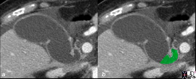
Neck of the Gallbladder |
| The neck of the gallbladder is sually quite difficult to image. In this CTscan of an elderly fenmale patient the gallbladder is dilated so its component parts are distorted and easierto visualise. The partial sigmoid shape of the neck is overlaid in green.
82382c03.8s 82382c03 85F gallbladder shape neck of the gallbladder cystic duct size age time distended dilated parts fundus body neck infundibulum cystic duct distended dilated small amount of fluid in the gallbladder fossa ascites CTscan copyright 2008 Courtesy Ashley Davidoff MD |
It is the most medial part of the gallbladder and lies in close association with the porta hepatis.
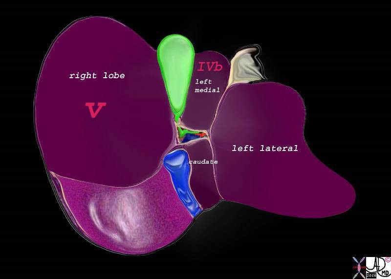
Relationship of the Neck to the Porta Hepatis |
|
The neck is the most posterior and medial portion of the gallbladder and is in close association with the porta hepatis which lies even more posterior and medial to it. 82220b01.2k05.82s liver gallbladder porta hepatis hepatic artery portal vein IVC inferior vena cava falciform ligament ligamentum teres bare area of the liver left lobe segment IV segment I caudate lobe quadrate lobe medial segment left ;obe lateral segment left lobe gastrohepatic ligament gallbladder right lobe hepatic Davidoff art copyright 2008 |
Applied Biology
The full extent of the neck is not routinely visualized on US because of its smal size, tortuosity and the fact that is nestled deep among other structures .
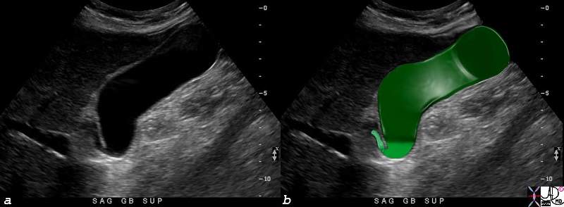
Parts of the Gallbladder – Ultrasound |
| The full extent of the neck of the gallbladder is not easily nor routinely seen on all ultrasound examinations because it is small, tortuous, and nestled among other echogenic structures that tend to camouflage it. In this instance, the neck (lightest green can be seen between the infundibulum and the beginning of the cystic duct.
82416c04.8s gallbladder parts fundus = darkest green body = dark green neck = light green cystic duct = lightest green normal anatomy USscan ultrasound Courtesy Ashley DAvidoff MD copyright 2008 |
As the structure with the smallest diameter and most tortuous course, it is the site that has a predilection to stone impaction.
Stone Impacted in the Neck Causing Obstruction |
|
The neck of the gallbladder is the narrowest and most tortuous portion of the gallbladder and thus there is a predilection for stones to become impacted and cause obstruction. If the obstruction is complicated by inflammation then acute cholecystitis ensues. 11921.8b05b019.8s gallbladder cystic duct gallstones cholelithiasis stone impacted in the cystic duct distended enlarged Davidoff Art copyright 2008 |
Hartmann’s pouch (aka ampulla of the gallbladder, fossa provesicalis, pelvis of the gallbladder) is a spheroidal or conical pouch at the junction of the neck with the cystic duct. It is a outpouching on the inferior surface of the neck that is thought to be acquired during life and caused by stone disease resulting in inflammation and secondary adhesions between the neck and the cystic duct.
References
Cystic Duct
Turner MA Fulcher AS The Cystic Duct: Normal Anatomy and Disease Processes Radiographics. 2001;21:3-22
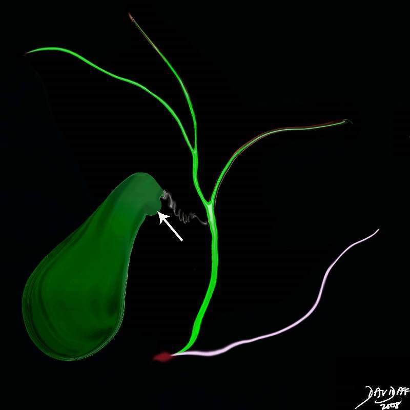
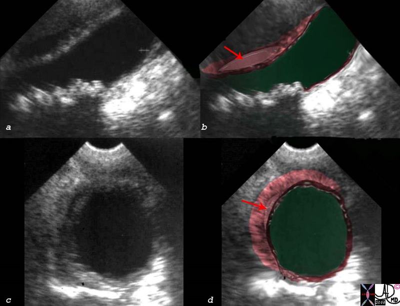
Hartmann’s Pouch |
| Hartmann’s pouch (arrow) is an outpouhcing situated on the inferior aspect of the neck of the gallbladder that is caused by prior inflammatory and stone disease in the area.
04766b05b07.32k.8s Hartmann’s pouch infundibulum anatomy stone disease acquired gallbladder Courtesy Ashley Davidoff MD copyright 2008 |