
copyright 2008
Definition
A thickened gallbladder is a radiological finding in which the wall is thicker than the normal 2-3mm.in the fasting state. It is normally only about 1mm thick in the neonate. Thickening of the gallbladder wall is always abnormal, although the significance of the thickening is highly dependent upon the underlying disease process.
It can be caused by multiple factors and conditions including renal failure, liver failure heart failure, ascites, hypoproteinemia, and inflammation. Other causes include carcinoma, HIV cholangiopathy, slceroderma. In carcinoma the thickening may be focal or diffuse. Thickening may also be due to the hyperplastic cholecystoses, such as adenomyomatosis or hypercholesterolosis. Adenomyomatosis gives a more characteristically focal thickening, while cholesterolosis may be more diffuse. In general, most pathological processes affecting the gallbladder will cause thickening, either due to inflammation or due to precipitation of bile contents due to prolonged stasis.
The diagnosis is made on imaging studies. Ultrasound and CT are both sensitive to the diagnosis.
Treatment strategies are based on the cause of the wall thickening
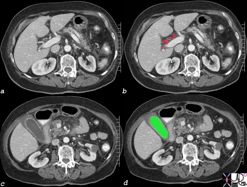
Edema – Cystic Arterioles in the Submucosa |
| The submucosal thickening of the gallbladder is characteristic of nonspecific edema
82006c04.8s liver gallbladder blood supply hepatic artery cystic artery cystic arterioles submucosa wall edema hyperemic mucosa red = hepatic artery green = lumen of the gallbladder CTscan Courtesy Ashley Davidoff MD Copyright 2008 |
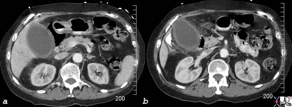
Acalculous Cholecystitis – RUQ Pain, Distended Gallbladder, Indurated Fat |
| Diffuse thickening with pericystic induration of the fat is most characteristic of inflammatory change. In this case perforated acalculous cholecystitis was the diagnosis.
77717c02s right upper quadrant pain ICU setting RUQ pain gallbladder distended thick walled hyperemic enhancing wall pericholecystic induration dirty fat stranding acalculous cholecystitis Courtesy Ashley DAvidoff MD copyright 2008 |
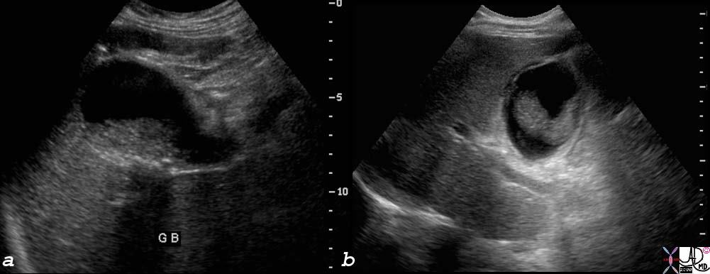
Acalculous Cholecystitis – RUQ Pain, Distended Gallbladder, Tumefactive Bile – same patient |
| Thickened bile (sludge) and fluid in the gallbladder fossa from the same patient above with acalculous cholecystitis.
77717c01s right upper quadrant pain ICU setting RUQ pain gallbladder distended thick walled sludge cholestasis tumefactive bile fluid in gallbladder fossa small shadowing stones acalculous cholecystitis shape of tumefactive bile in the gallbladder has fetus like formation USscan ultrasound Courtesy Ashley DAvidoff MD copyright 2008 |
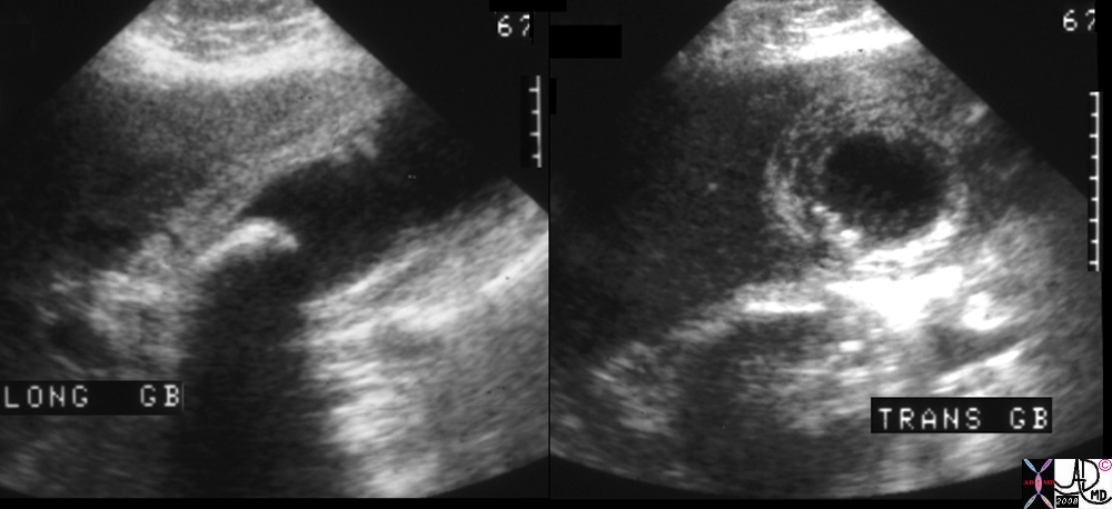
Acute Calculous Cholecystitis |
| Layering and thickening of the wall associated with pain and a stone is characteristic of calculous cholecystitis
00401.1cs 67Male right upper quadrant pain RUQ pain positive Murphy’s sign gallbladder gallbladder fossa thickened linear lacy thickening gall stones calculi calculous large stone in the infundibulum shadowing multiple small stones cholelithiasis cholecystitis acute cholecystitis acute calculous cholecystitis USscan ultrasound Courtesy Ashley Davidoff MD copyright 2008 |

Hepatitis |
| Diffuse thickening of the wall in a patient with acute hepatitis.
48008c01 gallbladder thick wall non distended dx hepatitis USscan Davidoff MD |
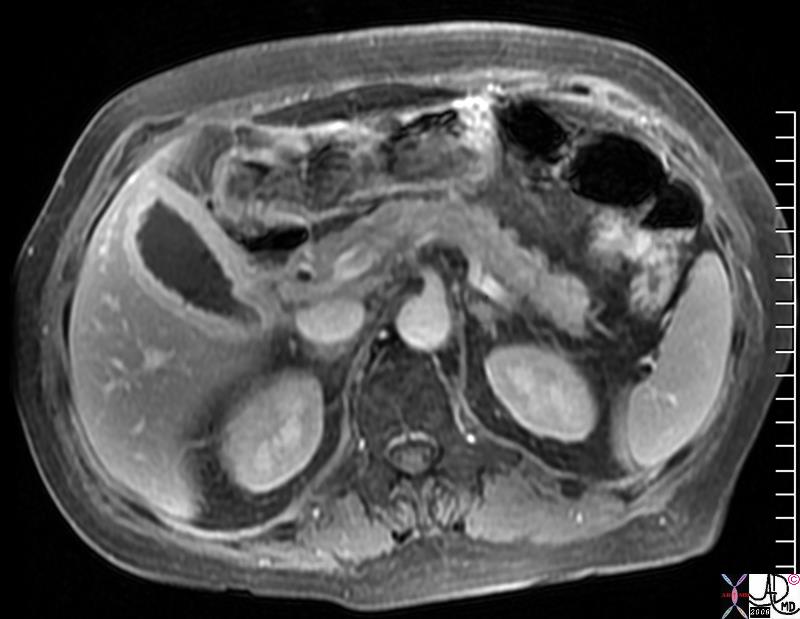
Acute on Chronic Cholecystitis |
| MRI showing hyperemia, stones, and thickening of the wall of the gallbladder in this patient with acute on chronic cholecystitis.
77718.8s.jpg gallbladder thick wall filling defects stones cholelithiasis chronic cholecystitis enhancing wall acute cholecystitis on chronic pancreas normal MRI fat suppression gadolinium C+ Courtesy Ashley Davidoff MD copyright 2008 |
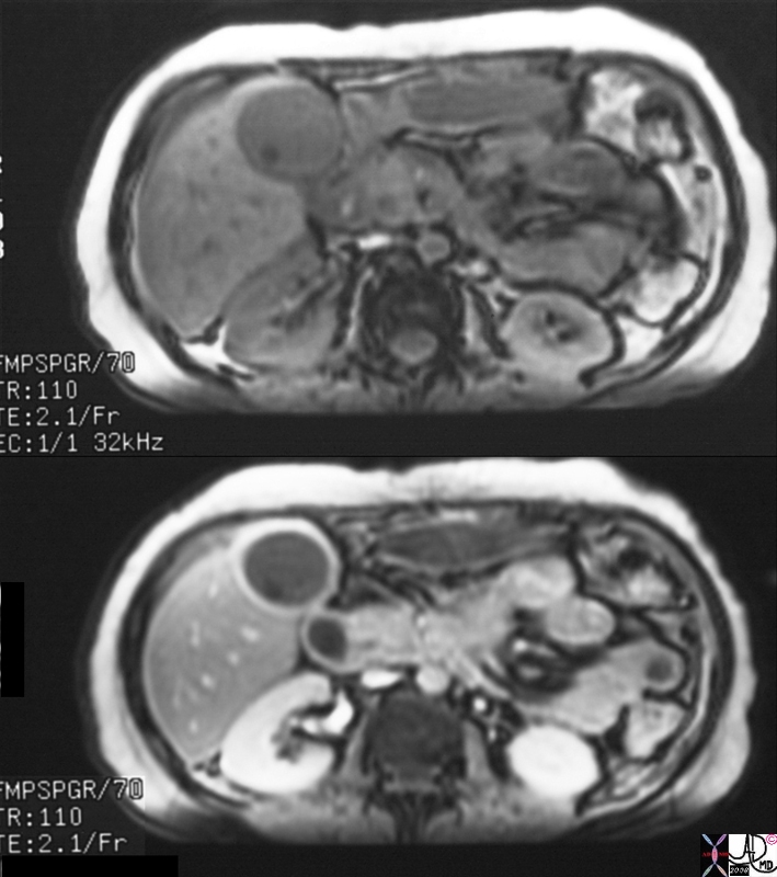
Chronic Cholecystitis Enhancing Wall |
| Thickening and hyperemia (pre and post contrast) of the wall and a single stone is shown on the MRI of a patient with chronic cholecystitis
16239c.8s gallbladder stone cholelithiasis wall thickened thick wall enhancing wall dx chronic cholecystitis bile duct cholangiocarcinoma MRI with and without contrast |
Chronic Cholecystitis -Tiny Calcified Cholesterol Floaters |
| Hyperemic wall and small cholesterol floaters – Chronic cholecystitis shown by CTscan
25306.8s small cholesterol crystals stones floaters hyperemic wall chronic cholecytitis gas air cholelithiasis CTscan Courtesy Ashley Davidoff MD copyright 2008 |
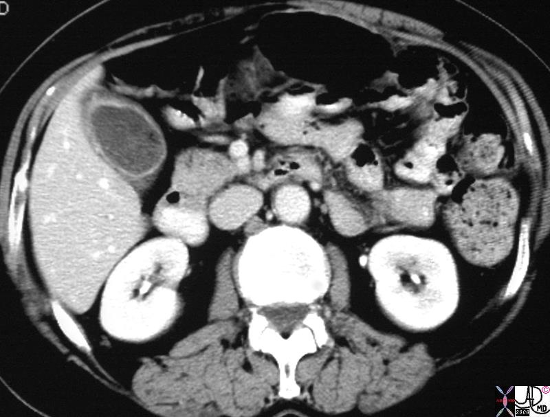
Chronic Cholecystitis, Hyperemic Wall, Small Contracted Gallbladder |
| Hyperemic wall and single moderate sized calcified stone – Chronic cholecystitis shown by CTscan
30701.8s stone cholelithiasis hyperemic wall small contracted chronic cholecytitis stomach gastrostomy redundant mucosa CTscan Courtesy Ashley Davidoff MD copyright 2008 chronic cholecytitis |
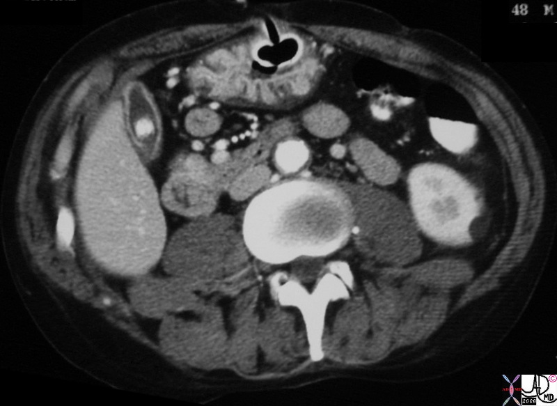
Chronic Cholecystitis Small Contracted Gallbladder with Stone and Thick Wall |
| Small contracted gallbladder with a thick wall – Chronic cholecystitis shown by CTscan
81916.8s gallbladder stone cholelithiasis thick wall thickened wall small contracted chronic cholecystitis CTscan Courtesy Ashley Davidoff MD copyright 2008 |
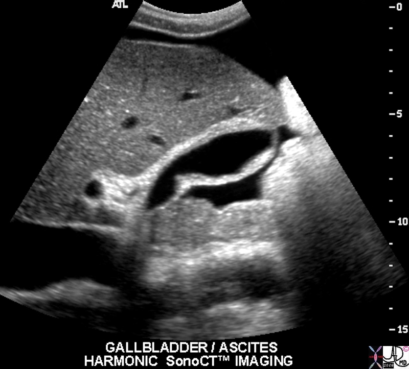
Ascites |
| Thick walled gallbladder in a patient with ascites shown by ultrasouns. The wall in this case is diffusely echogenic.
2005H5~1.8s abdomen abdominal cavity peritoneal cavity free fluid ascites gaallbladder thick walled ultrasound ascites USscan Courtesy Philips Medical Systems copyright 2008 |
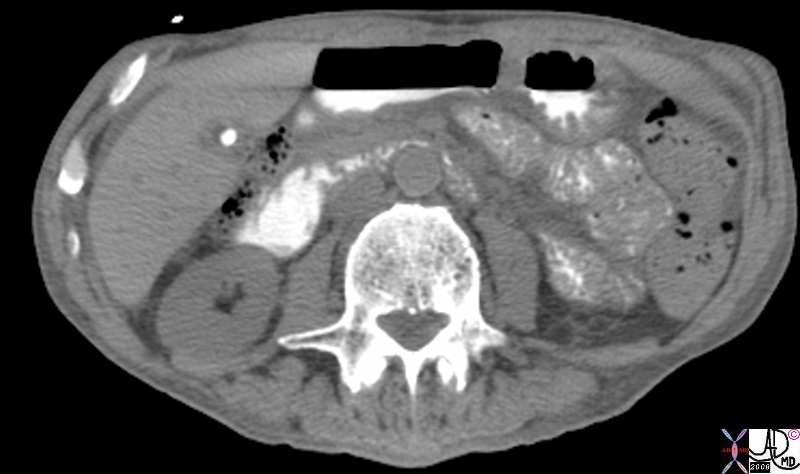
HIV Cholangiopathy Gallbladder fossa |
| HIV positive patient with known pancreatitis and a fever 41249c Courtesy Ashley Davidoff MD code pancreas pseudocysts code gallbladder fx fluid in the fossa imaging radiology CTscan inflammation infection(Image courtesy of Ashley Davidoff M.D.) |
Acute Right Heart Failure and Wall Edema |
| Acute tricuspid regurgitation shows an associated edematous wall of the gallbladder caused by congestion and poor lyphatic and venous drainage from the gallbladder. This entity can cause RUQ pain that simulateds acute cholecystitis. gallbladder lumen is not distended in this instance.
00425 gallbladder wall fx edema dx CHF liver heart cardiac dx CHF liver CHF acute cardiac failure CTscan Courtesy Ashley Davidoff MD copyright 2008 |
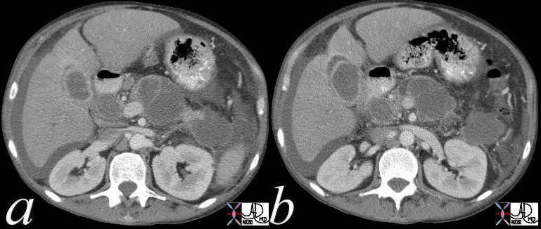
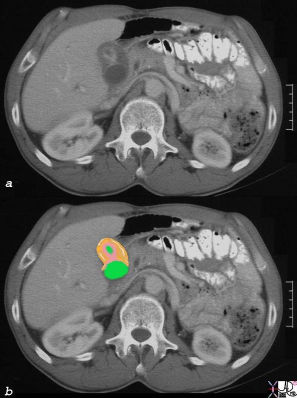
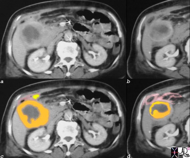
Liver Metastases – Edematous Galbladder Wall Due to Venous and Lymphatic Congestion |
| 51 year old male with history of rectal carcinoma with extensive hepatic metastases. (orange) The metastases surround gallbladder and by space occupation result in venous and lymphatic congestion causing a thickened edematous gallbladder wall (orange) with a small lumen (green) Tthe portal triad shows surrounding edema (white arrow). This is called periportal tracking which is another sign of venous and lymphatic engorgement. The metastases are calcified confirming the mucinous nature of the tumor.
calcified metastasis in gallbladder fossa edema of the wall lymphatics contracted gallbladder CTscan copyright 2008 Courtesy Ashley Davidoff MD gallbladder liver portal triad 82313c01.8s |
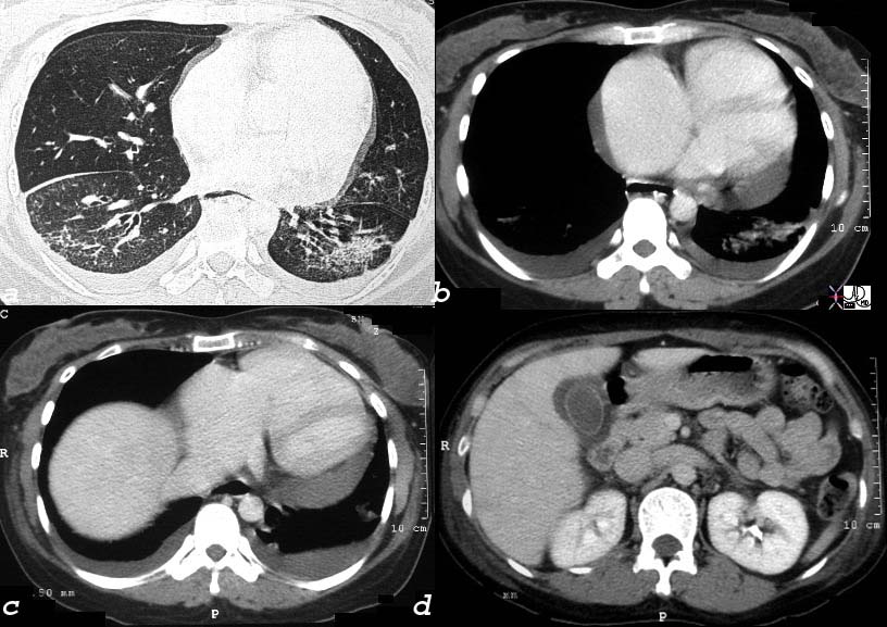
Scleroderma Pulmonary Hypertension and Right Heart Failure |
| 30464c02 32 female lungs pleura heart cardiac RA RV right ventricle right atrium pericardium gallbladder esophagus ILD basal interstitial lung disease pericardial effusion pleural effusion cardiomegaly enlarged esophagus patulous esophagus gallbladder wall edema congestive cardiac failure RHF right heart failure pulmonary hypertension cor pulmonale dx scleroderma CTscan Courtesy Ashley Davidoff MD |
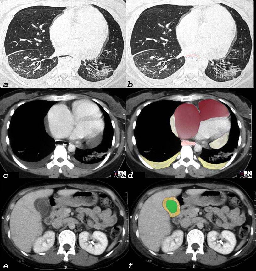
Scleroderma Pulmonary Hypertension and Right Heart Failure |
|
This 32 year old female has scleroderma and her disease is characterized by interstitial basal lung disease (a,b) with normal anterior lung fields and fibrotic posterior lung fields, enlargement of right heart structures (maroon in d), pericardial effusion (light yellow in d), pleural effusions (dark yellow in d) and an edematous wall (orange, in f) of the gallbladder (lumen is green). As a result of her lung disease she developed pulmonary hypertension, right heart failure and tricuspid regurgitation and this lead to the gallbladder edema. and 30464c08 32 female lungs pleura heart cardiac RA RV right ventricle right atrium pericardium gallbladder esophagus ILD basal interstitial lung disease pericardial effusion pleural effusion cardiomegaly enlarged esophagus patulous esophagus gallbladder wall edema congestive cardiac failure RHF right heart failure pulmonary hypertension cor pulmonale dx scleroderma CTscan Courtesy Ashley Davidoff MD |

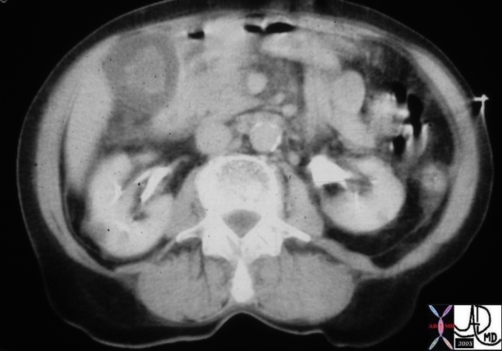
Empyema – Complex Fluid Filled Gallbladder |
| Empyema of the gallbladder and thickened inflammed wall.
48010.800 gallbladder fx thick walled shadowing cholelithiasis complex fluid dx gallbladder empyema Courtesy Laura Feldman |
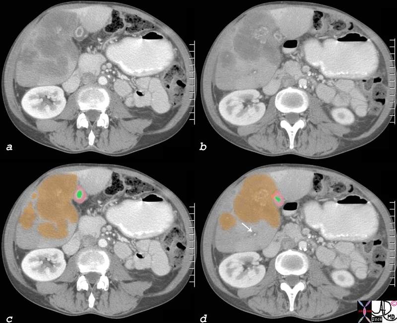
Carcinoma of the Fundus Mucosal and Submucosal Infiltration |
| Focal thickening of the gallbladder fundus in this patient with gallbladder carcinoma.
16228c01.8s gallbladder wall submucosal tumor = orange mucosal tumor = pink lumen = green thickened mucosa and submucosa carcinoma of the gallbladder CTscan Courtesy Ashley DAvidoff MD copyright 2008 |
Gallbladder Carcinoma with Perforation and Abscess Formation |
| Diffuse irregular thickening of the gallbladder wall in patient with gallbladder carcinoma.
16254c01b02.8s gallbladder thickened irregular wall air anterior wall small fluid collection gallbladder carcinoma complicated by perforation and abscess formation CTscan Courtesy Ashley Davidoff copyright 2008 |
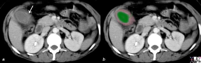
Gallbladder Laceration from Trauma |
| The 32 year old male suffered from a motor vehicle accident and presented with right upper quadrant pain. The gallbladder shows disruption of the mucosal enhancement (arrow in a) and is thick walled (pink) These findings are consistent with a a laceratio and possible rupture of the gallbladder.
16216c01.8s 32 male s/p trauma MVA gallbladder hyperemic mucosa thickened wall disrupted mucosa decompresssed lumen CTscan Courtesy Ashley Davidoff MD copyright 2008 |
References
Gallbladder Wall Thickening Web presentation from radiologyassistant.nl 5star
Van Breda Vriesman et al AJR