
CT scanning of the Gallbladder
Ashley Davidoff MD Alok Anand
Definition
CT scan is the a radiological study that uses X-ray technology combined with computer technology to visualize the structures of the body. In general for abdominal imaging, it provides a global view of all the abdominal structures, providing excellent results in almost all patients since there is little operator dependance. Both intravenous and oral contrast are utilized, and the current multidetector technology enables the whole abdomen to be scanned in 10-15 seconds. With respect to the gallbladder no specific protocols are used and the intent of using CT is to visualize structures related to the gallbladder disease, and not necessarily the gallbladder itself, which is best visualized by ultrasound.
Principles
Computed tomography is an imaging technique where X-ray technology and computer technology are used to produce transverse or axial tomograms of the body. X-ray generators and detectors are positioned in a doughnut type ring around the body. The X-ray generators produce highly collimated X-ray beams which interact with the tissue from many different angles and the result is detected and recorded by the detectors that are aligned in a ring oppposite the generators. Each tissue has specific attenuation characteristics. The attenuation of the x-rays of a specific tissue obtained from multiple projections is collected by a computer which organizes and displays the information using mathematical algorithms.
Indications
CT provides a global view of the abdomen and is indicated as secondary study for most gallbladder disease particularly when complications are suspected. In acute cholecytitis when peritonitis or gallbladder perforation is suspected CT enables the visualization of the surrounding tissues particulalrly mesenteric fat which is not appprecuated by ultrasound or HIDA scan. Merizzi’s syndrome when cholecystitis and bile duct obstruction are both present and caused by a stone impacted in the infundibulum causes bile duct obstruction is best evaluated by CT since shadowing caused by ultrasound may preclude accurate diagnosis. The characterization of hemorrhage in acute hemorrhagic cholecystitis is also not appreciated by ultrasound but tthe presence of this entity does not have any therapeutic significance other than the blood clot could be the cause of obstruction. In the setting of blunt trauma to the abdomen with suspected gallbladder injury the ability to acquire a global view of the abdomen provided by CT scan is essential. When gallbladder ileus is suspected then a global view of the gallbladder, biliary system, and small bowel, which is best provided by CT scan. Gallbladder carcinoma that requires staging is best performed with PET CT.
Contraindications
There is no absolute contraindication to utuilizing CT since it its benefits almost always outweigh the risks. The only signicant real contraindication is using CT during the first trimester, when crucial development is taking place.
The radiation created by CTs is ionizing is potentially carcinogenic, but may also cause mutations and birth defects. In patients who are of reproductive age and unable to verify pregnancy status, a pregnancy test should always be performed first.
CT should also not be used in young children if ultrasound has potential appl;ication
Contrast studies should be exercised with caution in the patient populations mentioned above, as well as in patients with renal insufficiency, as iodinated contrast can cause further damage to renal parenchyma, possibly resulting in acute kidney injury requiring dialysis, or even death.
Advantages
CT imaging offers a global view of the abdomen. With one or two sequences it allows us to see the organs, the surrounding tissues, the gastrointestinal tract, small calcifications, peritoneal fat and mesenteries, blood vessels and the bones mostly with exquisite detail. CT can be used to image both soft-tissue and bony tissue. It is particularly useful for assessing free air in the gallbladder and nearby structures. As imaging technologies have improved, it is now commonly able to visualize the gallbladder and nearby structures within the span of one breath hold. Thus fine structures, such as the biliary tree can be well visualized.
CT can be used during interventional procedures, allowing percutaneous drainage, biopsy or ostomy placement,avoiding more invasive therapies.
Disadvantages
In addition to exposing patients to potentially harmful ionizing radiation it carries risk of radiation exposure to healthcare personnel as well. As such, its use should be accompanied by appropriate safety precautions, such as adequate shielding and the use of failsafe techniques. Personnel should also be monitored regularly for cumulative exposure to ionizing radiation.
Aim
The purpose of using CT scan in suspected or known gallbladder disease is to get a sense of the extent of disease that may have occurred beyond the gallbladder confines.
Method
Patient Preparation
The patient should be fasting, and both intravenous and oral contrast is utilized.
Equipment
The gallbladder and its surrounds do not move and as long as the patient can hold their breath, any of the modern MDCT scanners are able to provide information needed
Technique
Typically gallbladder imaging is done with a fasting patient in supine position . Both intravenous and oral contrast are utilized. The patient then passes through the gantry, as the source spins around it, passing x-rays through at multiple angles, to be detected by sensors within the gantry and sent to a computer for processing and reconstruction. Once generated, these images may be further processed to reconstruct images in slices along other planes, such as coronal, sagital, or oblique.
Result
When gallbladder disease is suspected ultrasound is the study of choice. CT is used as a secondary study to evaluate for the spread of disease. The sensitivity and specificity of CT in diagnosing gallbladder pathologies depends heavily on the suspected pathology. It is highly sensitive for acute calculous cholecystitis, gangrene, tumors, and hemorrhage.
Examples of Various Diseases
Fat and Air
 Subtle Character Changes Subtle Character Changes |
| 70269c02 gallbladder air methane nitrogen gas cholesterol stone isodense with bile fat subtle change CTscan |
Porcelain Gallbladder
 Porcelain Gallbladder Porcelain Gallbladder |
| The gallbladder in this instance is contracted, and has porcelain like calcification. This suggests chronic cholecytitis though no stones are identified. The patient requires to undergo elective cholecystectomy.
47683c01 gallbladder fx calcification in wall calcified wall dx porcelain gallbladder CTscan Davidoff MD |
Abscess
Early Abscess Formation |
|
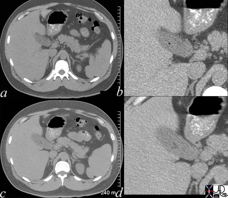
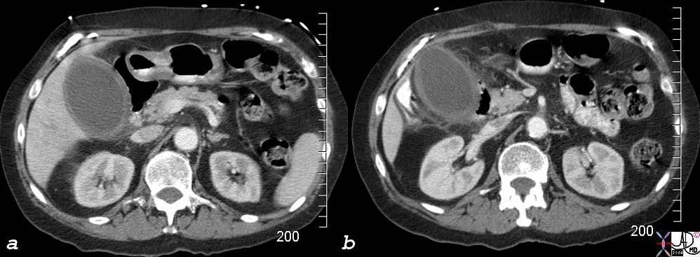
The CTscan is from a patient who has a fever and right upper quadrant pain and who has acute cholecystitis. A small fluid collection is seen in the gallbladder fossain both images a and b that reflects early abscess formation. 16188c.8s gallbladder hyperemic thickened wall early abscess formation fluid collections in the gallbladder fossa acute cholecystitis CTscan Courtesy Ashley Davidoff MD copyright 2008 |
Early Abscess Formation |
|
The CT scan through the gallbladder shows a second case of early spread of the infection into the gallbladder fossa. The yellow more linear collections represent the early purulence which at this stage are too small for drainage and a trial of antibiotics would be the treatment of choice. 16188c05.8 gallbladder hyperemic thickened wall early abscess formation fluid collections in the gallbladder fossa acute cholecystitis CTscan Courtesy Ashley Davidoff MD copyright 2008 |

Abscess in the Gallbladder Fossa |
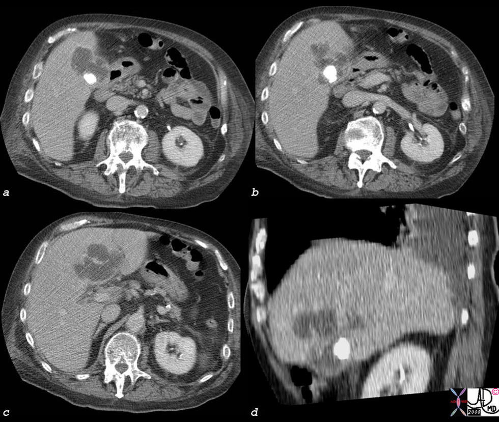
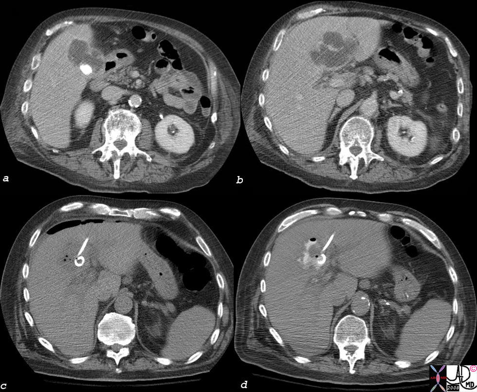
| The abscess in the gallbladder fossa in this patient is large and irregular and is associated with a large calcified stone in the gallbladder. Images a,b,c, are in the transverse plain, while image d is in the coronal plane. It is in the latter situation, where the stone can be seen in the relatively small gallbladder and the abscess in the gallbladder fossa and within the liver is optimally seen. percutaneous drainage and antibiotics is indictated. In this instance gallbladder carcinoima should alsd be considered in the appropriate clinical setting.
44364c02s elderly patient with fever right upper quadrant pain, RUQ pain, elevated white cell count gallbladder cholelithiasis acute cholecystitis fluid in the gallbladder fossa gallbladder abscess liver abscess hepatic abscess CTscan ‘Courtesy Ashley Davidoff MD copyright 2008 |
Abscess in the Gallbladder Fossa Treated with CT Guided Catheter Drainage |
|
The patient above was treated with percutaneous drainage, (c) was decompressed overnight and the abscessogram performed the following day shows contrast in a decompressed abscess. Drainage should continue till the cavity is less than 5-100ccs in volume and the drainage is less than 5-10ccs over 24 hours. 44364c03s elderly patient with fever right upper quadrant pain, RUQ pain, elevated white cell count gallbladder cholelithiasis acute cholecystitis fluid in the gallbladder fossa gallbladder abscess liver abscess hepatic abscess percutaneous drainage catheter drainage MIT minimally invasive therapy CTscan ‘Courtesy Ashley Davidoff MD copyright 2008 |
Ruptured Acalculous Cholecytitis
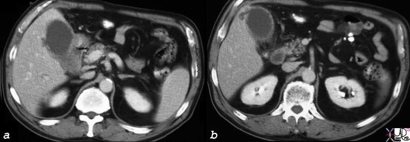
Acalculous Cholecystitis – RUQ Pain, Distended Gallbladder, Indurated Fat |
| 77717c02s right upper quadrant pain ICU setting RUQ pain gallbladder distended thick walled hyperemic enhancing wall pericholecystic induration dirty fat stranding acalculous cholecystitis Courtesy Ashley DAvidoff MD copyright 2008 |
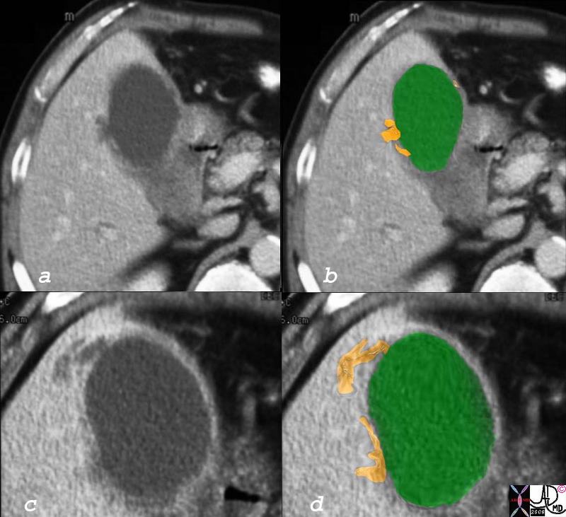
Direct Invasion into the Liver and Bile Duct Obstruction |
|
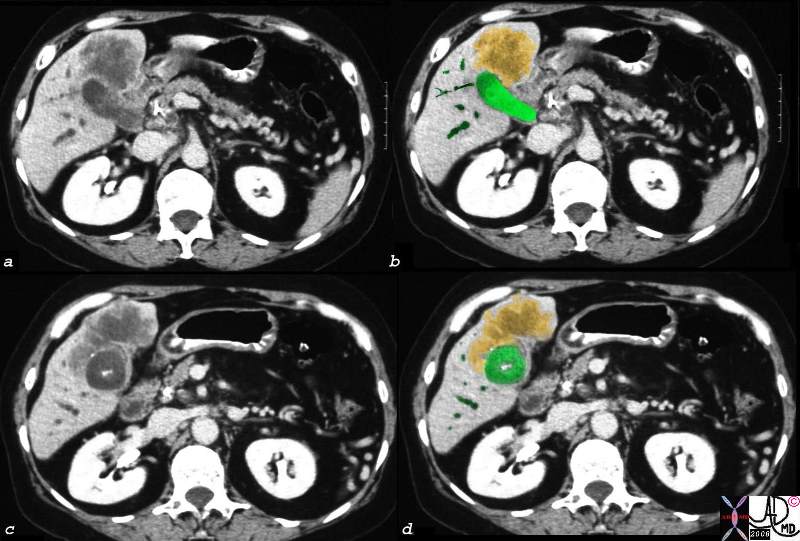
The CTscan of this patient shows a normal sized gallbladder (green) associated with a 4-5cms mass (orange) adjacent to the gallbladder, and extending from the gallbladder fossa. There is asssociated biliary obstruction (dark green tubes) This case represents invasive gallbladder carcinoma with bile duct obstruction. Images c and d show the almost universal association of gallstones in patients with carcinoma. In this case the stones (white) are in the centre of the gallbladder. (green) 16254c03.8s gallbladder anterior wall liver invasion space occupatopn obstruction bile ducts aggressive gallbladder carcinoma complicated by direct invasion metastasis liver windows narroe windws tumor settings gallbladder fossa GBF CTscan Courtesy Ashley Davidoff copyright 2008 |
Contrast reaction varying from mild to life threatening anaphylaxis can occur. Contrast extravasation into the subcutaneous tissues also occurs occasionally.
CT has its largest application in gallbladder disease as a secondary study, where the primary diagnosis has usually been established by ultrasound or HIDA scan and the extent of disease is in question. Evaluation for ruptured gallbladder and extent and staging of gallbladder cancer are two examples.