
The Cystic Artery
The gallbladder is supplied by the cystic artery. The cystic artery is usually a branch of the right hepatic artery but can also originate from left or common hepatic arteries, the gastroduodenal artery, or directly from the celiac trunk.
The Cystic Artery Anterior and Posterior Cystic Arteries off the Right Hepatic Artery |
| gallbladder artery hepatic artery right hepatic artery normal anatomy angiogram angiography cystic artery branches Courtesy Ashley Davidoff copyright 2012 Courtesy Ashley Davidoff 05222c02.8s |
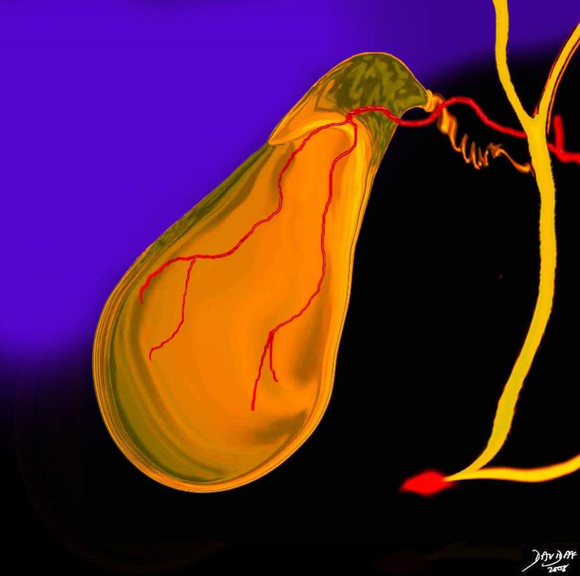
The cystic artery passes superior to the cystic duct and terminates in two branches called the anterior (superficial) branch that runs on the free wall of the gallbladder and the posterior (deep) branch that runs on hepatic aspect of the gallbladder close to the gallbladder fossa. The cystic duct is supplied by 2 to 4 minor branches called Calot’s arteries that originate just proximal to the origin of the anterior and posterior vessels.
Cystic Artery off the Right Hepatic Artery |
| Copyright 2012 Courtesy Ashley Davidoff MD 04766b05b04.55k.8s |
The vessel for most of its course runs in the triangle of Calot whose boundaries are the inferior border of the liver edge, cystic duct, and common hepatic duct.
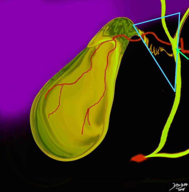
Calot’s Triangle |
|
Calot’s triangle is an anatomic space bordered by the common hepatic duct medially, the cystic duct inferiorly and the liver superiorly. The cystic artery usually passes through the triangle, and Calot’s lymph node is also within this triangle. It is an important landmark to recognize during laparoscopic cholecystectomy gallbladder Copyright 2012 Courtesy Ashley Davidoff 04766b05b04.57dk.8s |
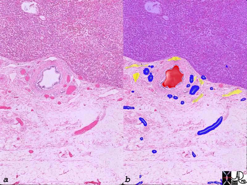
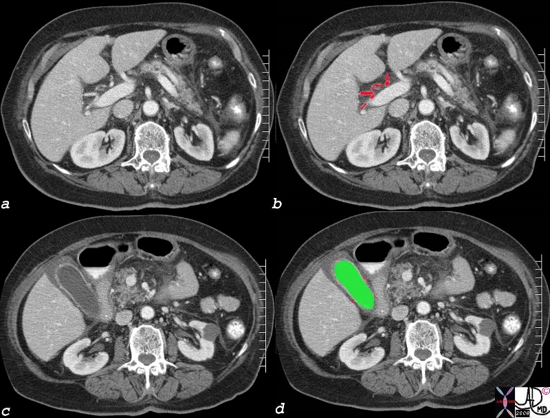
Small arteries, veins and biliary radicles run between the liver and the gallbladder in the gallbladder fossa directly from the liver into the gallbladder.
Arteries, Veins, and Lymphatics in the Gallbladder Fossa |
|
The histological section of the gallbladder fossa shows the relatively large thick walled branch of the deep cystic artery, (red) abutting the liver (upper portion purple) accompanied by venules (blue) and lymphatics. (yellow) The veins drain directly into the fossa and connect with intrahepatic portal branches of the segment V and IV of the liver. gallbladder gallbladder fossa gbf venules arterioles liver connective tissue hilum of the gallbladder venous drainage blood supply Copyright 2012 Courtesy Ashley Davidoff MD 00140c03.8s |
Cystic Arterioles in the Submucosa Deep and Superficial Branches |
|
Image shows the hepatic artery in the porta hepatis, while c and d, show the tiny vessels in the lamina propria (red dots) of the gallbladder since the edema in the wall has provided sufficient contrast to expose them. Courtesy Ashley Davidoff MD Copyright 2008 82006c04.8s
|
Variations
In about 15% of patients the anterior and posterior arteries have separate origins and this entity is called double cystic artery. An example is seen below.
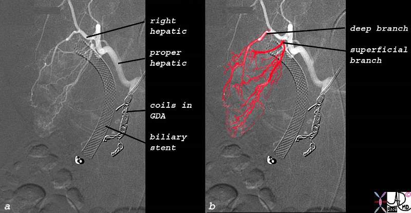
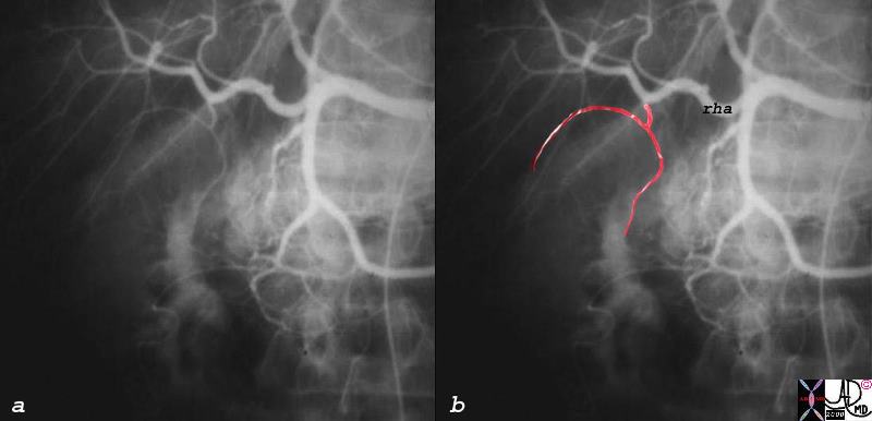
Double Cystic Artery – Separate Origins Off the Right Hepatic Artery |
| This is the angiogram of a 52 year old male with HIV cholangiopathy and cryptosporidium associated with a neoplastic mass in the CBD, and a fistula between the CBD and duodenum presents with uncontrolled hemobilia. Embolization of the gastroduodenal artery was successful. The follow up angiogram shows the catheter in the GDA which has been occluded by coils, a stent in the CBD, and flow in the cystic circulation.
The right hepatic artery gives rise to separate origins of the deep or posterior branch that runs close to the gallbladder fossa and the superficial or anterior branch that runs on the free wall of the gallbladder. Copyright 2012 Courtesy David Lee MD 78240c01.8s 52 |
Other variations of the origin of the vessel include its origin from the left or or common hepatic artery, and it can course either anterior to or posterior to the common hepatic duct
Historical Aspects.
The Vesalius drawing of the exposed gallbladder is a remarkable rendition of vessels and ducts traveling to and from the gallbladder.
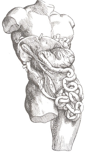
The Art of Vesalius |
| 13237 abdomen small bowel liver gallbladder anatomy Vesalius historical drawing DB |
portal vein.